Figure 4. Pharmacological dissection of retinal circuits conveying signals from photosensitized bipolar cells. A: Example trial bin count (TBC) of light-responsive cell exposed to a 2 s light pulse (irradiance 1013 photons/cm2/s) starting at time 0 s, with the progressive addition of drugs blocking glutamatergic signaling (trial order runs from bottom
to top, with timing of the addition for individual agents indicated by the horizontal lines to left; note that once added
the agent was included until the end of the trial, such that for this example the recording ended with artificial cerebrospinal
fluid (aCSF), meclofenamic acid (MFA), ((S)-1-(2-amino-2-carboxyethyl)-3-(2-carboxy-5-phenylthiophene-3-yl-methyl)-5-methylpyrimidine-2,4-dione
(ACET), L-AP4, and 6,7-dinitroquinoxaline-2,3-dione (DNQX) all included in the recording media. B: Example TBC of light-responsive cell exposed to a 2 s light pulse (irradiance 1013 photons/cm2/s) at starting at time 0 s under the progressive addition of agents affecting the gamma-aminobutyric acid (GABA)ergic blockade
(protocol and depiction as in A). C: A model of inner retinal circuitry following ectopic expression of rhodopsin in ON bipolar cells consistent with the pharmacological
manipulations reported here. MFA blocks gap junctions, inhibiting propagation of oscillations originating in AII amacrine
cells (ACs). MFA is also expected to block the transfer of signals from ON rod bipolar cells to ON cone bipolar cells via
AII ACs but not transfer from ON rod bipolar cells to OFF cone bipolar cells or more direct signaling from ON cone bipolar
cells. Inclusion of an inhibitory AC between the ON cone bipolar cell and the ON RGC allows for sign inversion to produce
excitatory responses to light and accounts for the impact of the GABAergic blockade (B). CBCs= cone bipolar cells; RGCs = retinal ganglion cells; TPMPA = (1,2,5,6-tetrahydropyridin-4-yl)methylphosphinic acid.
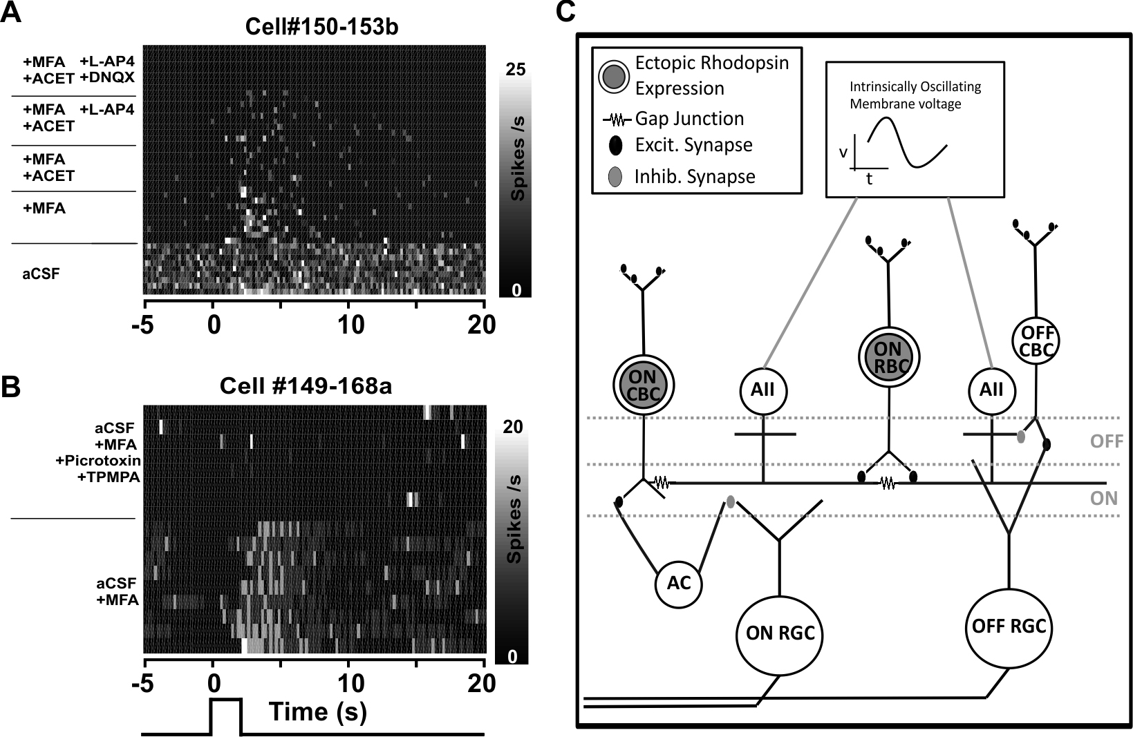
 Figure 4 of
Eleftheriou, Mol Vis 2017; 23:334-345.
Figure 4 of
Eleftheriou, Mol Vis 2017; 23:334-345.