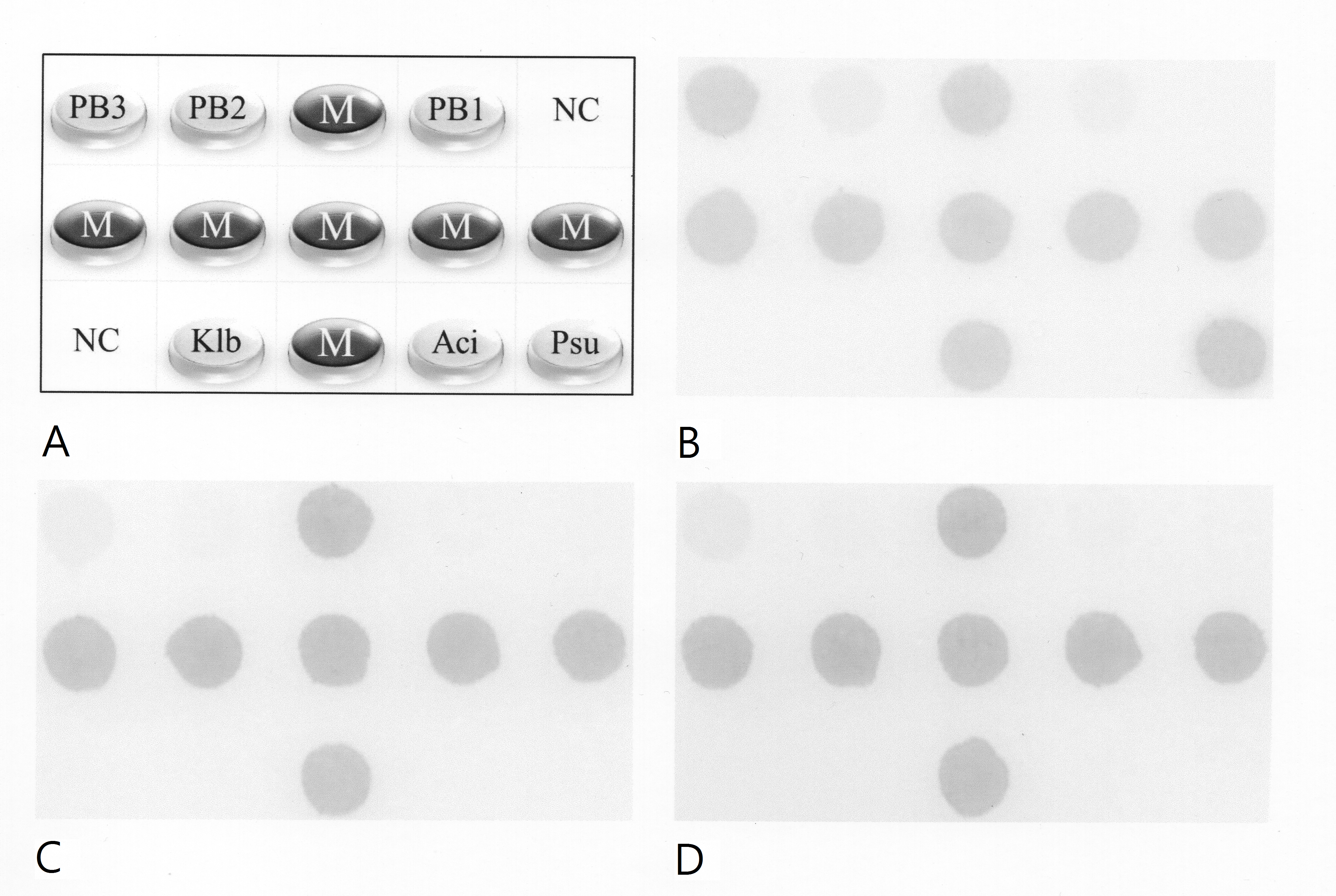
Figure 1. The bacterial dot hybridization assay.
A: Layout of oligonucleotide probes on the array (0.6 cm × 0.4 cm). The universal bacteria probes PB1, PB2, and PB3 were designed
from a conserved region at the 3′ end of the 16S rRNA gene. Dots labeled “NC” were negative controls (tracking dye only).
Dots labeled “M” were position markers, that is, an irrelevant digoxigenin-labeled oligonucleotide. Dots labeled “Psu,” “Aci,”
and “Klb” were genus-specific probes used to identify the
Pseudomonas,
Acinetobacter, and
Klebsiella species, respectively. Probe sequences for all dots were shown in our previous study [
34].
B−
D: Representative hybridization patterns for 10 ng, 10 pg, and 10 fg of
P. aeruginosa DNA.
 Figure 1 of
Fang, Mol Vis 2017; 23:306-317.
Figure 1 of
Fang, Mol Vis 2017; 23:306-317.