Figure 3. Analysis of the antiviral, zinc oxide tetrapods in mitigating HSV-1(KOS)tk12 infection in HCE cells. A: Scanning electron microscope image of zinc oxide tetrapod nanoparticles. B: Representative bright-field microscopic images of infected and zinc oxide-treated infected HCE cells to assess viral entry
via the 5-bromo-4-chloro-3-indolyl-β-D-galactoside (X-gal) substrate. C: Quantification of viral entry and spread assessed by amount of blue present via Metamorph software. Significance was determined
by t-test: p<0.0001. D: Representative immunoblot image (left panel) and its quantification (right panel) for HSV-1 gB from cell lysates of untereated
and zinc oxide-treated infected HCE cells. Note: data used for the immunoblot analysis and quantification of cell lysates
in panel D were taken from HCE cells infected with the wild strain herpes simplex virus type-1 (HSV-1)KOS virus.
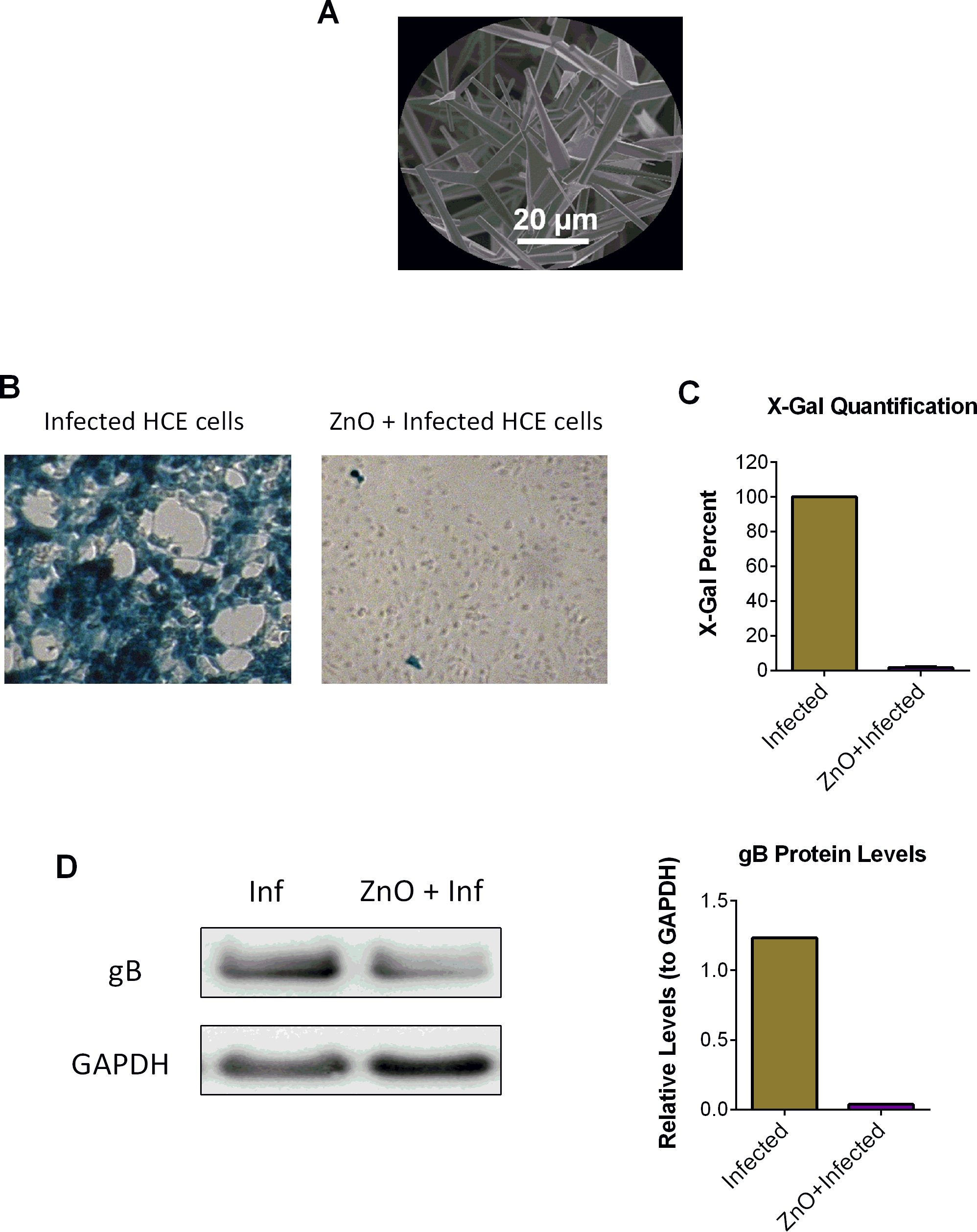
 Figure 3 of
Duggal, Mol Vis 2017; 23:26-38.
Figure 3 of
Duggal, Mol Vis 2017; 23:26-38.