Figure 2. Development of ex vivo pig corneal model for HSV-1(KOS)tk12 infection. A: General schematic of major steps undertaken to develop pig cornea cultures harvested from pigs aged 180-200 days. B: Representative images taken via a modular stereo microscope of uninfected and infected cornea treated with the 5-bromo-4-chloro-3-indolyl-β-D-galactoside
(X-gal) substrate. C: Representative HSV-1 gB immunoblot (left panel) and intensity quantification (right panel) present in uninfected and infected
corneal epithelial cell lysates.
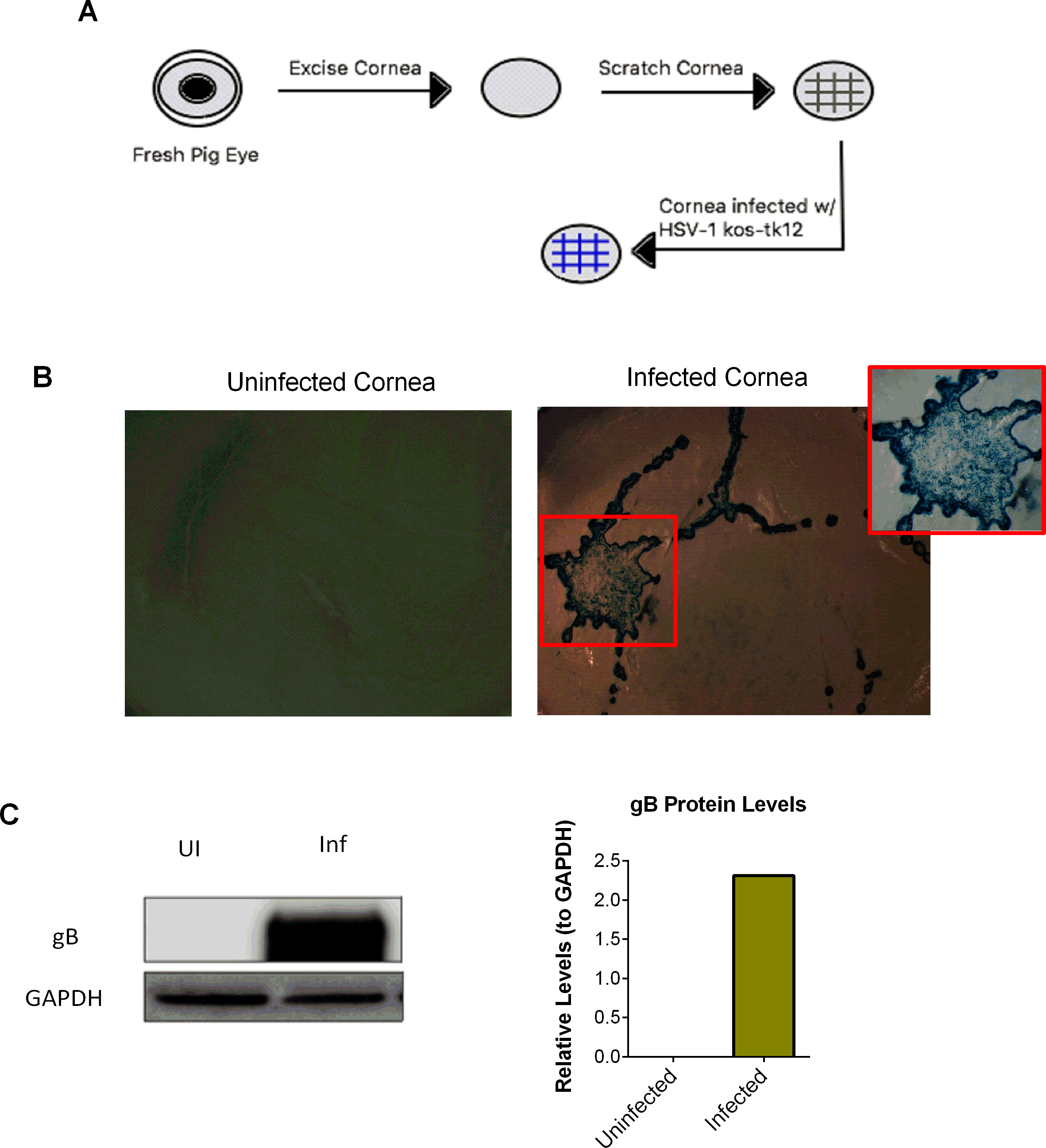
 Figure 2 of
Duggal, Mol Vis 2017; 23:26-38.
Figure 2 of
Duggal, Mol Vis 2017; 23:26-38.