Figure 1. Infection of HCE cells with the HSV-1(KOS)tk12 virus. A: Viral entry assay using the 5-bromo-4-chloro-3-indolyl-β-D-galactoside (X-gal) substrate to confirm entry. Viral entry is
confirmed by the prevalent blue observed in the infected human corneal epithelail (HCE) cells (blue, right panel). B: A representative immunoblot of the cell lysates for HSV-1 glycoproteins, gB and gD in uninfected and infected HCE cells.
C: Quantification of gD and gB viral protein levels of representative immunoblot.
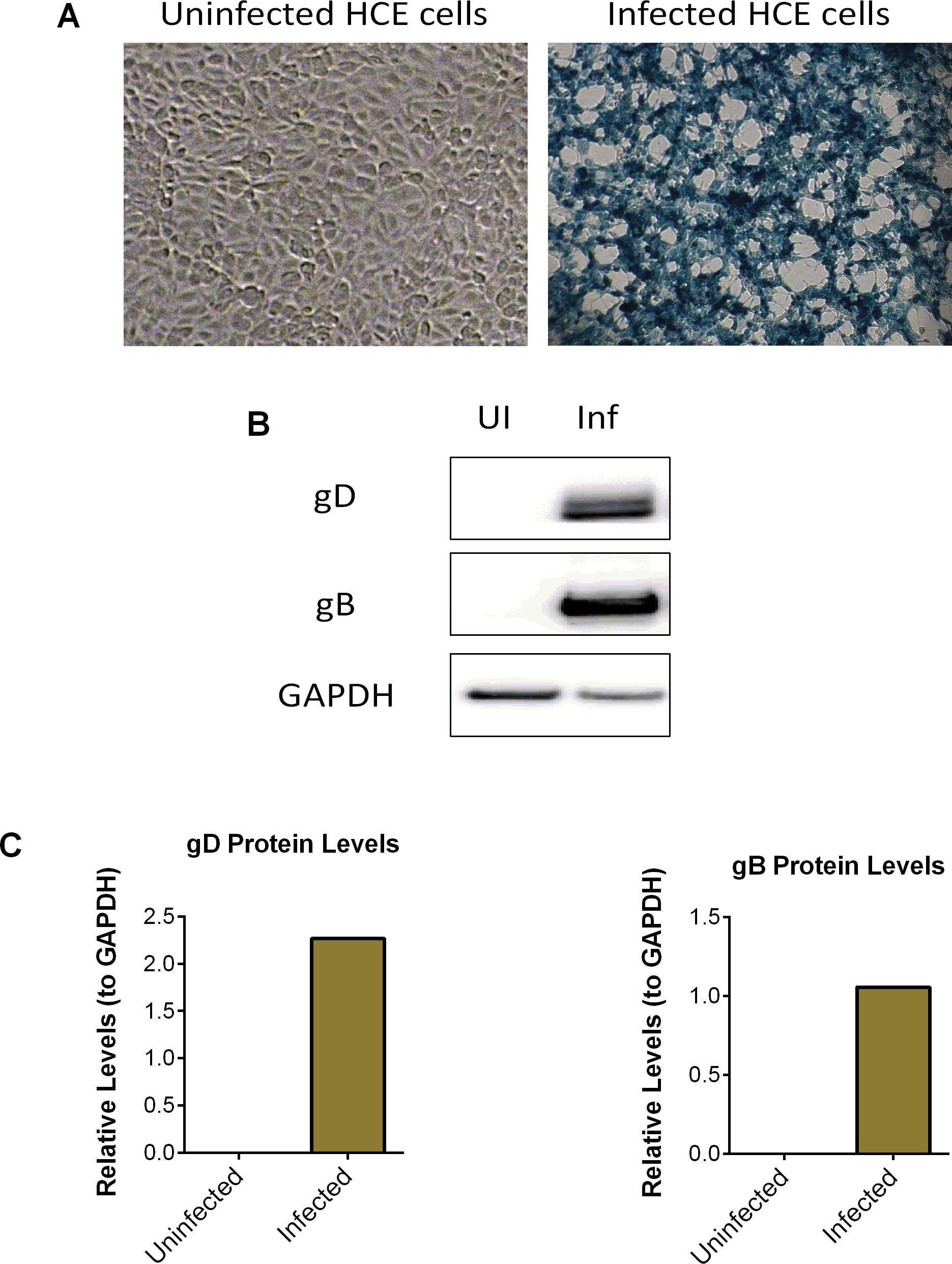
 Figure 1 of
Duggal, Mol Vis 2017; 23:26-38.
Figure 1 of
Duggal, Mol Vis 2017; 23:26-38.