Figure 1. Pathological observation in retinas of rats with STZ-induced DR. Representative photomicrographs of hematoxylin and eosin
(H&E) staining of retinopathy. Scale bars = 20 μm. A: The control group (n = 4) showed a homogenous surface. The inner and outer nuclear layers were all regularly arranged. B: In the group treated with streptozotocin (STZ; n = 4), morphological changes in the rats with diabetic retinography (DR)
were observed, including disruption and irregular arrangement of cells in the outer and inner nuclear layers. C: The vehicle group (n = 4) showed a disordered structure and irregular cells in the outer and inner nuclear layers. D: The resolvin D1 (RvD1) group (n = 4) illustrated that the inner and outer nuclear layers were tighter and the cells were
more regular. Original magnification = 400X. GCL = ganglion cell layer; INL = inner nuclear layer; ONL = outer nuclear layer.
E: The permeability of the blood–retinal barrier was tested with the Evans blue permeation assay. The leakage in the retinas
of the diabetic rats after 3 months was statistically significantly higher than that in the control group. **p<0.01. Values
are expressed as mean ± standard deviation (SD); n = 4 experiments.
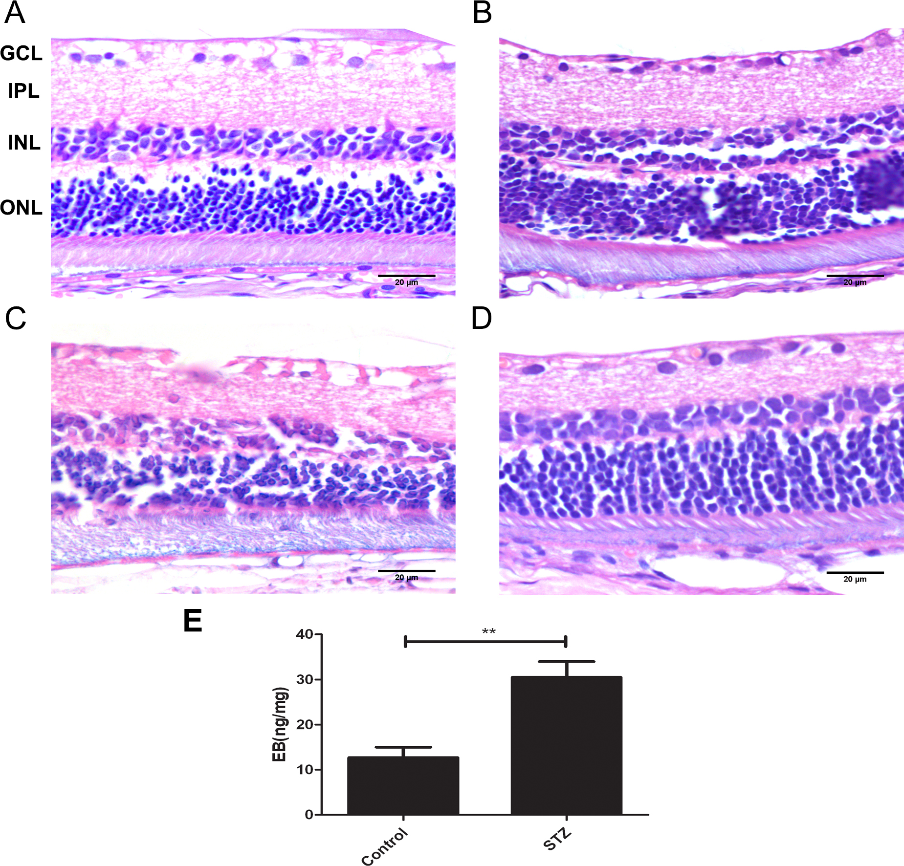
 Figure 1 of
Yin, Mol Vis 2017; 23:242-250.
Figure 1 of
Yin, Mol Vis 2017; 23:242-250.