Figure 9. CNV in aged Atg5ΔRPE and Atg7ΔRPE mice. Representative electronic micrographs showing the RPE area of 17-month-old wild-type (+/+), Atg5ΔRPE (Atg5−/−), and Atg7ΔRPE (Atg7−/−) mice. Note the blood vessels (thin arrows) in the RPE layer of the Atg5ΔRPE or Atg7ΔRPE mice. Thick arrow indicates a capillary endothelial cell (Atg5−/−) or a platelet (Atg7−/−). Scale bar = 5 µm.
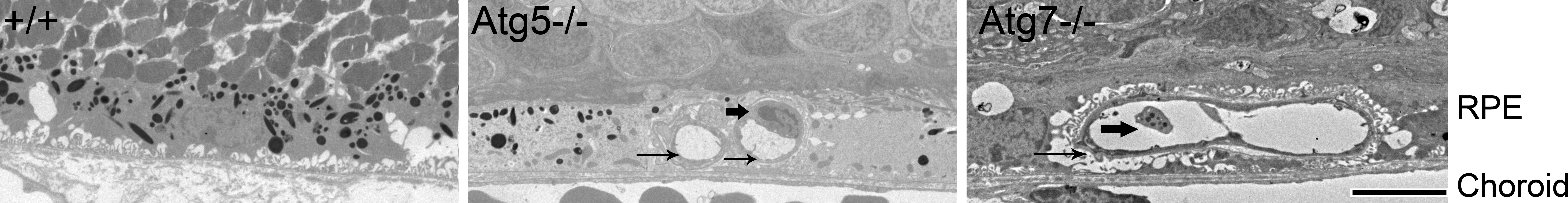
 Figure 9 of
Zhang, Mol Vis 2017; 23:228-241.
Figure 9 of
Zhang, Mol Vis 2017; 23:228-241.