Figure 7. Thinned RPE in aged Atg5ΔRPE and Atg7ΔRPE mice. Representative electronic micrographs showing the RPE region of the 17-month-old wild-type (+/+), Atg5ΔRPE (Atg5−/−), and Atg7ΔRPE (Atg7−/−) mice. Note that the RPE thicknesses of the Atg5ΔRPE and Atg7ΔRPE mice are approximately one third to one half that of the wild-type mice. The wild-type and Atg5ΔRPE mice shown were pigmented, and the Atg7ΔRPE mouse was albino. Arrow indicates a hypotrophic RPE cell. Scale bar = 10 µm.
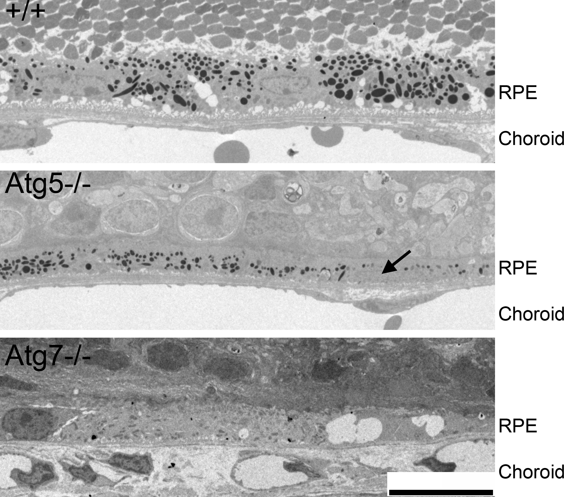
 Figure 7 of
Zhang, Mol Vis 2017; 23:228-241.
Figure 7 of
Zhang, Mol Vis 2017; 23:228-241.