Figure 5. Retinal degeneration in aged Atg5ΔRPE and Atg7ΔRPE mice. Representative images of toluidine blue–stained sections showing the retina layers of 17-month-old wild-type, Atg5ΔRPE, and Atg7ΔRPE mice. Note the retina layers of the Atg5ΔRPE and Atg7ΔRPE mice without retinal degeneration (no rd) were similar to those of the wild-type mice but the outer plexiform layer (OPL),
ONL, IS, and OS were diminished in the Atg5ΔRPE and Atg7ΔRPE mice with retinal degeneration (rd). The image for the Atg7ΔRPE mice without retinal degeneration was from a pigmented background, and the other images were from albino mice. Arrows indicate
engorged RPE cells. GCL, ganglion cell layer; IPL, inner plexiform layer; INL, inner nuclear layer; OPL, outer plexiform layer.
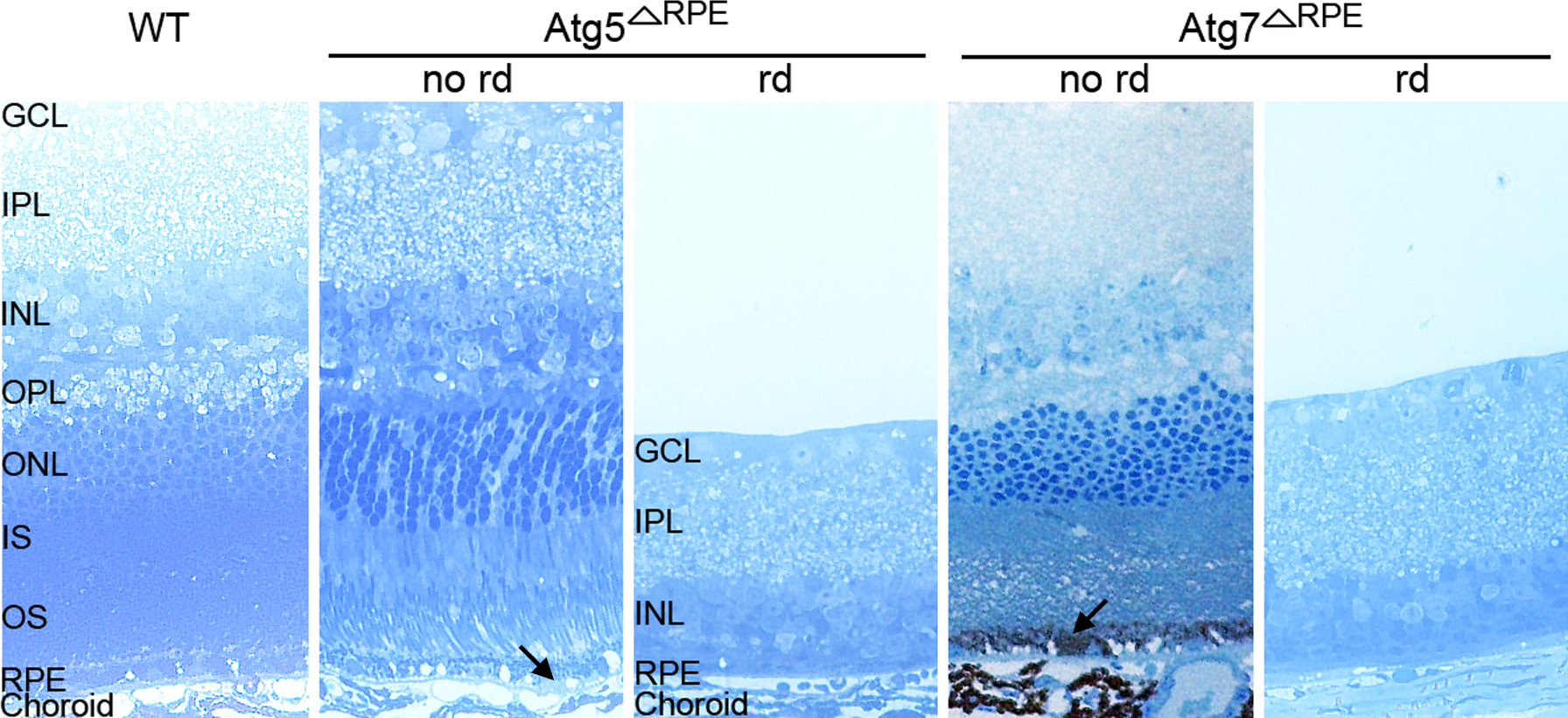
 Figure 5 of
Zhang, Mol Vis 2017; 23:228-241.
Figure 5 of
Zhang, Mol Vis 2017; 23:228-241.