Figure 1. RPE-specific deletion of Atg5 or Atg7 and p62/SQSTM1 accumulation in the RPE of Atg5ΔRPE and Atg7ΔRPE mice. A: Reverse transcription polymerase chain reaction (RT-PCR) showed that Atg5 or Atg7 was not expressed in RPE cells isolated from Atg5ΔRPE or Atg7ΔRPE mice but was expressed in the neuroretina isolated from these mice. RT–PCR using Efemp1 primers confirmed the integrity of the RNA from all the samples. B: Frozen sections from 8-month-old wild-type, Atg5ΔRPE, and Atg7ΔRPE mice were stained with an antibody (green signal) against p62/SQSTM1. Note the bright green staining in the RPE of the Atg5ΔRPE and Atg7ΔRPE mice. The nuclei were stained with 4’,6-diamidino-2-phenylindole (DAPI; blue signal). C: The p62 staining in the RPE was quantified using Image J software. n = 3 mice per genotype. Error bars indicate the mean
± standard deviation (SD). WT, wild-type; ONL, outer nuclear layer; IS, photoreceptor inner segment; OS, photoreceptor outer
segment.
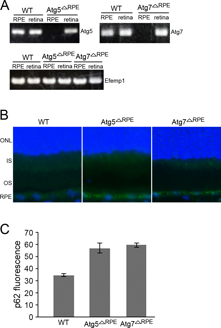
 Figure 1 of
Zhang, Mol Vis 2017; 23:228-241.
Figure 1 of
Zhang, Mol Vis 2017; 23:228-241.