Figure 4. Contribution of MMP-9 protein produced by infiltrating leukocytes to uveitic inflammatory injury after UVR. Both lenses, Etafilcon
A (right eye; A-E) and Nelfilcon A (left eye; F-J) contralaterally performed evaluation in a same mice to investigate the relative damage and protection after UVR. The arrowhead
in H indicate the hyperemic iris. The arrow in I and J indicate the infiltrating leukocytes. K-R: The expression of MMP-9 protein was found in the infiltrating leukocytes of corneal stroma (P), anterior chamber (L, N), and iridocorneal angle (P, R) after UVR. S: The protein levels of MMP-9 was evaluated by western blotting. T: Quantitative analysis of MMP-9 protein in the anterior segment of eye (n=4 per group). Scale bars: 25 μm. The p<0.01 indicated
the statistically significant.
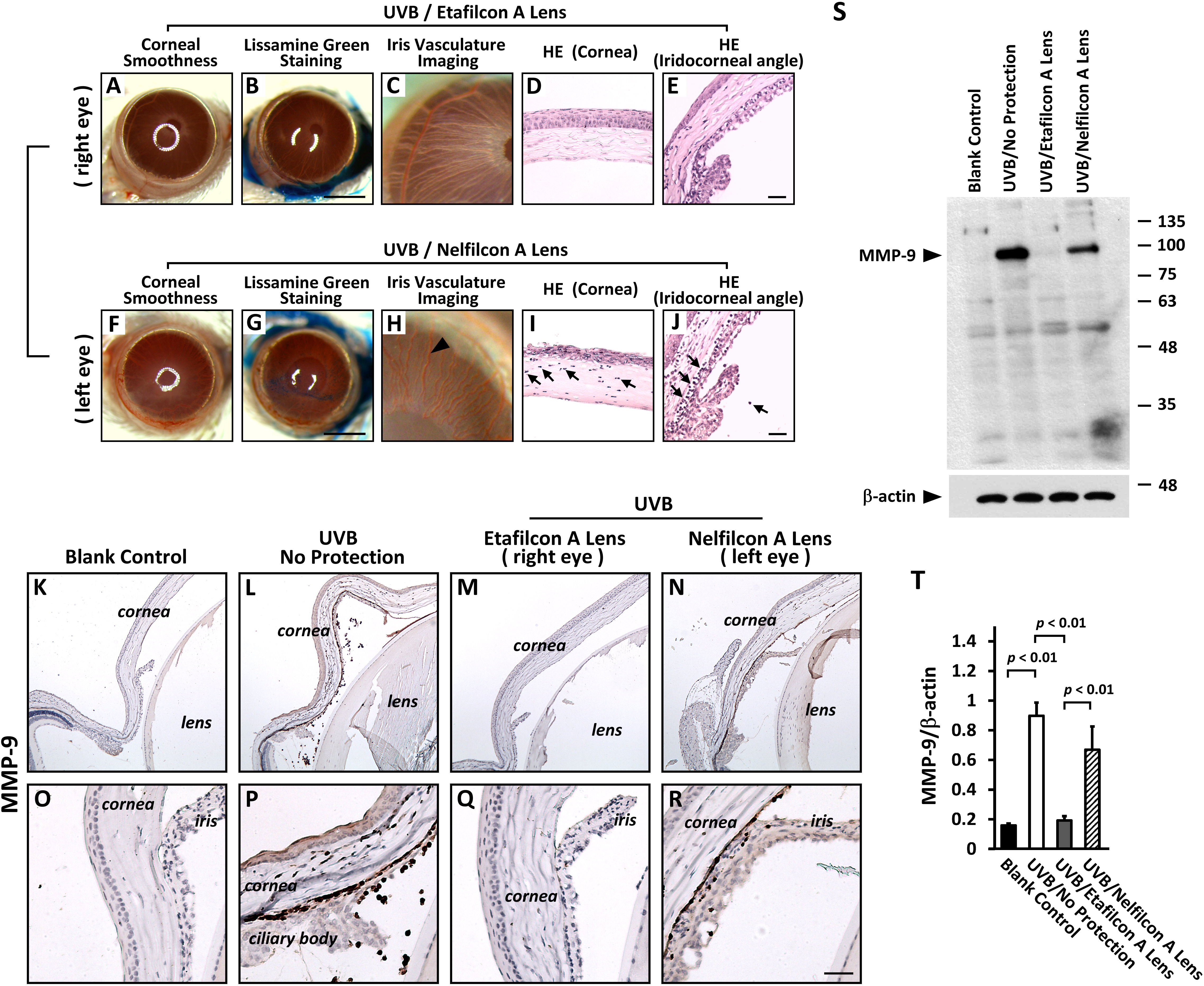
 Figure 4 of
Shao, Mol Vis 2017; 23:219-227.
Figure 4 of
Shao, Mol Vis 2017; 23:219-227.