Figure 3. UVR contribution to the pathogenesis of cellular infiltrates in the iridocorneal angle of eyes. A-H: Histological analysis of anterior eye. B, C: The influx of PMNs in the corneal stroma (indicated by arrow), and (F, G) iridocorneal angle (indicated by arrow). I: Quantitative analysis of PMNs in the corneal stroma (n = 11 per group). J: The incidence of corneal inflammatory injury (n = 11 per group). K: Quantitative analysis of PMNs in the iridocorneal angle (n = 11 per group). L: The incidence of uveitic inflammatory injury (n = 11 per group). M, N: Scatterplots indicated a significant correlation between the reduction in UVR strength and PMN recruitment inhibition. Scale
bars: 25 μm. The p<0.05 and p<0.01 indicated the statistically significant.
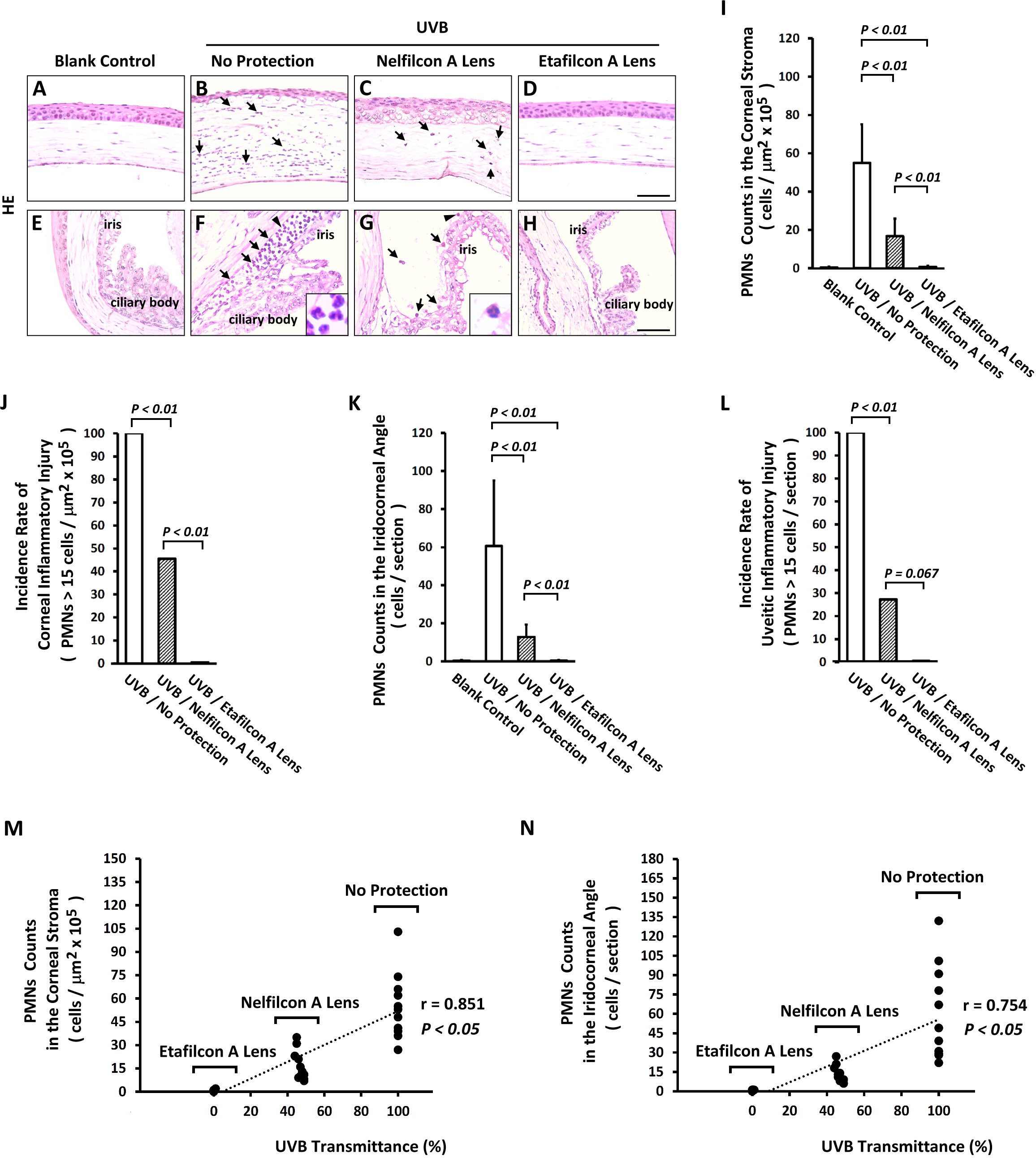
 Figure 3 of
Shao, Mol Vis 2017; 23:219-227.
Figure 3 of
Shao, Mol Vis 2017; 23:219-227.