Figure 2. UVR role in the pathogenesis of corneal and uveitic injury in anterior eye segments. The clinical corneal evaluation of (A-D) corneal smoothness, and (E-H) lissamine green staining. I: Quantitative analysis of corneal smoothness (n = 12 per group). J: The incidence of corneal smoothness (score>2). K: Quantitative analysis of lissamine green staining (n = 12 per group). L: The incidence of corneal staining (score >2). M-P: The clinical evaluation of anterior iris surface. N,O: The hyperemic change in the vessel on the anterior iris surface (arrowhead) and limbus (arrow). Q: Quantitative analysis of the clinical uveitis score (n = 12 per group). R: Quantitative analysis of the aqueous humor protein concentration (n = 7 per group). All scale bars = 1.25 mm. p<0.05; p<0.01.
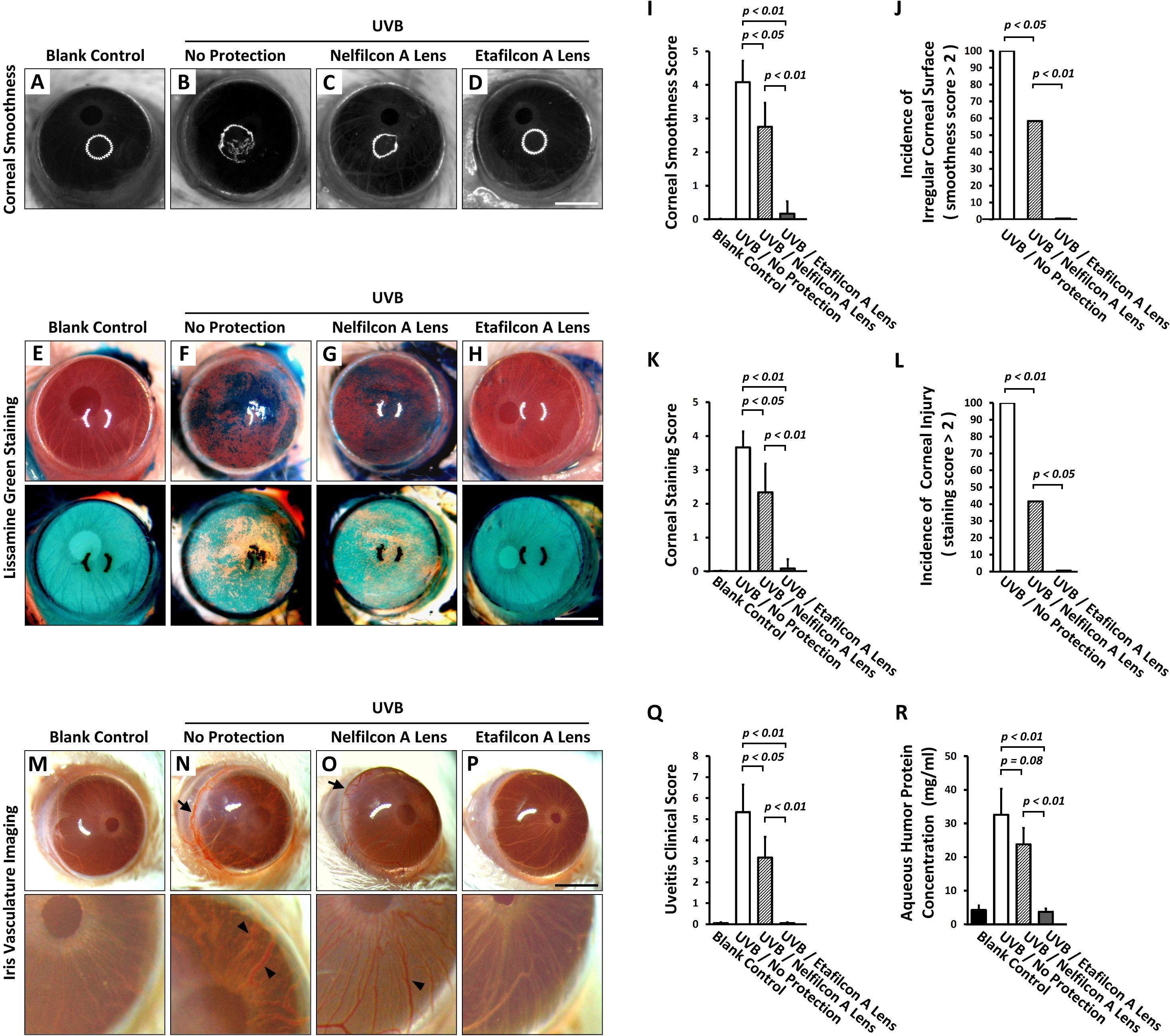
 Figure 2 of
Shao, Mol Vis 2017; 23:219-227.
Figure 2 of
Shao, Mol Vis 2017; 23:219-227.