Figure 4. Fluorescence photomicrographs and quantification of Iba1-labeled retinal microglia/macrophages following light damage. A: Longitudinal branched (green arrowheads) and amoeboid-shaped (white arrowheads) microglia/macrophages are shown in different
zones of the wild-type (WT) retina. B: Counting of Iba1-positive microglia/macrophages revealed increased numbers in the light-damaged retinas compared with the
non-light-damaged retinas. There were significantly fewer Iba1-positive microglia/macrophages in all zones of the C5aR knockout
(KO) retinas compared to the WT and C3aR KO retinas. n = 4, *p<0.05. GCL = ganglion cell layer, INL = inner nuclear layer,
ONL = outer nuclear layer, RPE = retinal pigment epithelium. C: Relative mRNA levels of Iba1 measured with real-time quantitative PCR (qPCR). No statistically significant differences were
observed in the mRNA levels of Iba1 among the non-light-damaged retinas of the WT, C3aR KO, and C5aR KO mice. There were statistically
significantly lower mRNA levels of Iba1 in the C5aR KO retinas compared with the WT and C3aR KO retinas after light damage.
n = 4, *p<0.05.
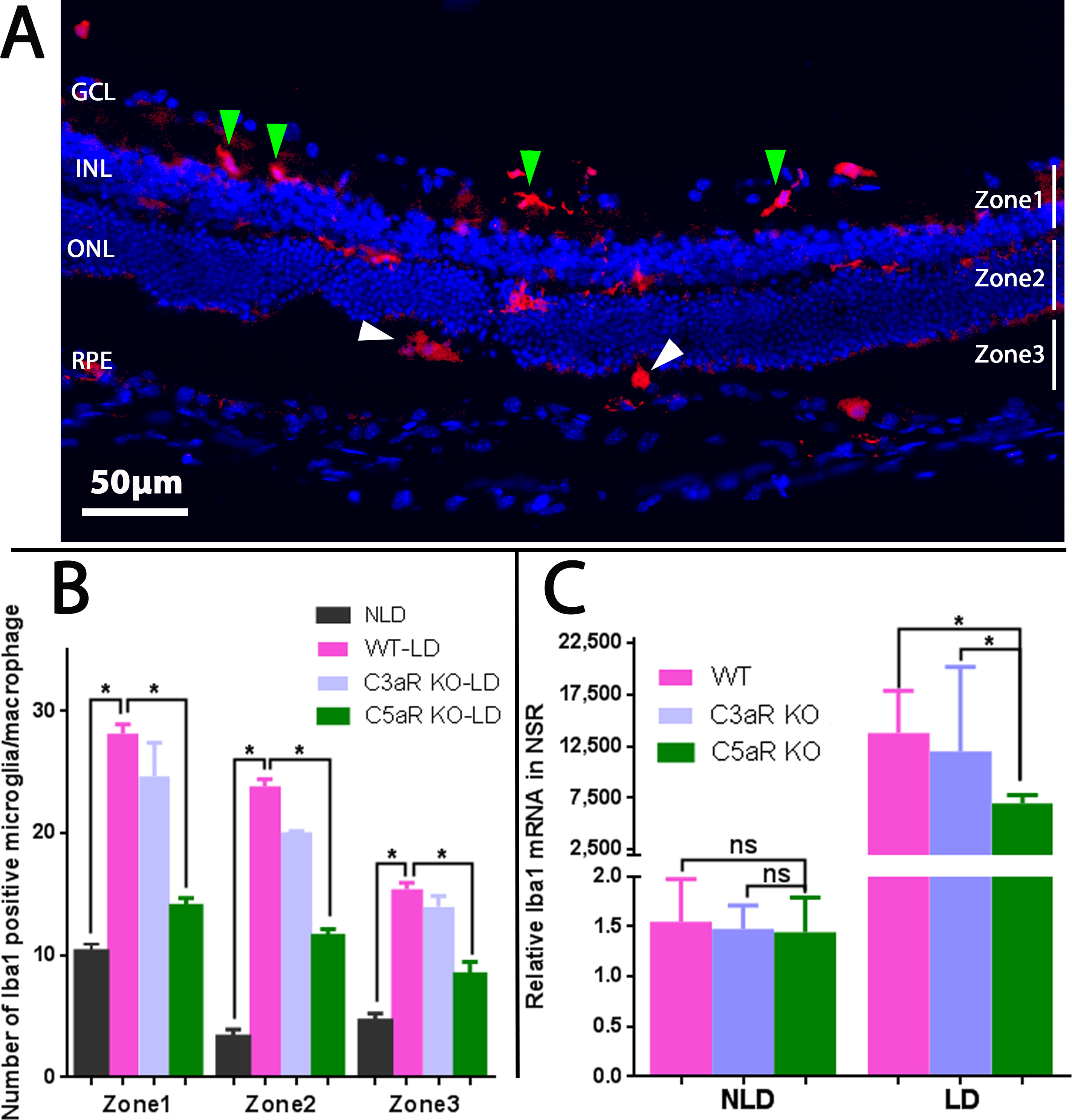
 Figure 4 of
Song, Mol Vis 2017; 23:210-218.
Figure 4 of
Song, Mol Vis 2017; 23:210-218.