Figure 3. Triple immunolabeling for Iba1, C3aR, and C5aR. Fluorescence photomicrographs show Iba1 (red), C3aR (green), and C5aR (dark
gray) in the retinas at day 2 after light damage. Retinal sections from the C3aR knockout (KO) mice and the C5aR KO mice were
used as controls for antibody specificity. Staining of the wild-type (WT) retinal sections with (first row) and without (second
row) primary antibody are shown. The merged images show colocalization of Iba1, C3aR, and C5aR (white arrows). GCL = ganglion
cell layer, INL = inner nuclear layer, ONL = outer nuclear layer, RPE = retinal pigment epithelium. Scale bar equals 50 μm.
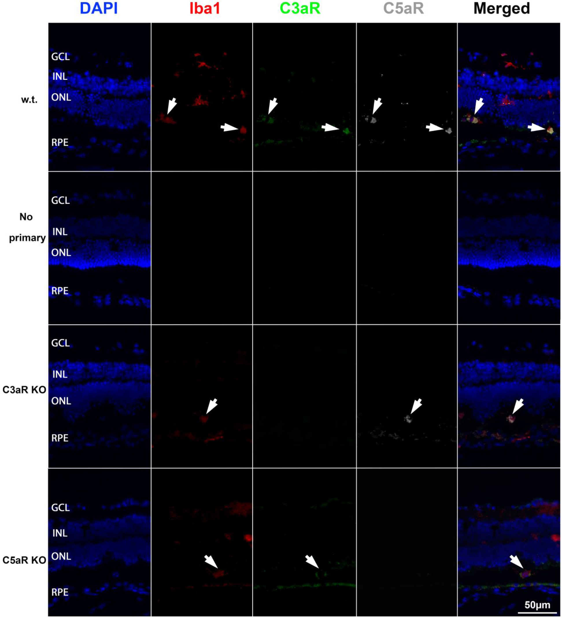
 Figure 3 of
Song, Mol Vis 2017; 23:210-218.
Figure 3 of
Song, Mol Vis 2017; 23:210-218.