Figure 2. Double immunolabeling for Iba1 and TSPO. Fluorescence photomicrographs show Iba1 (red) and TSPO (green) in the retinas at
day 2 after light damage. Retinal sections from wild-type (WT) mice labeled with secondary antibody served as a negative control
(upper row). The merged images show colocalization of Iba1 and TSPO (white arrows). GCL = ganglion cell layer, INL = inner
nuclear layer, ONL = outer nuclear layer, RPE = retinal pigment epithelium. Scale bar equals 50 μm.
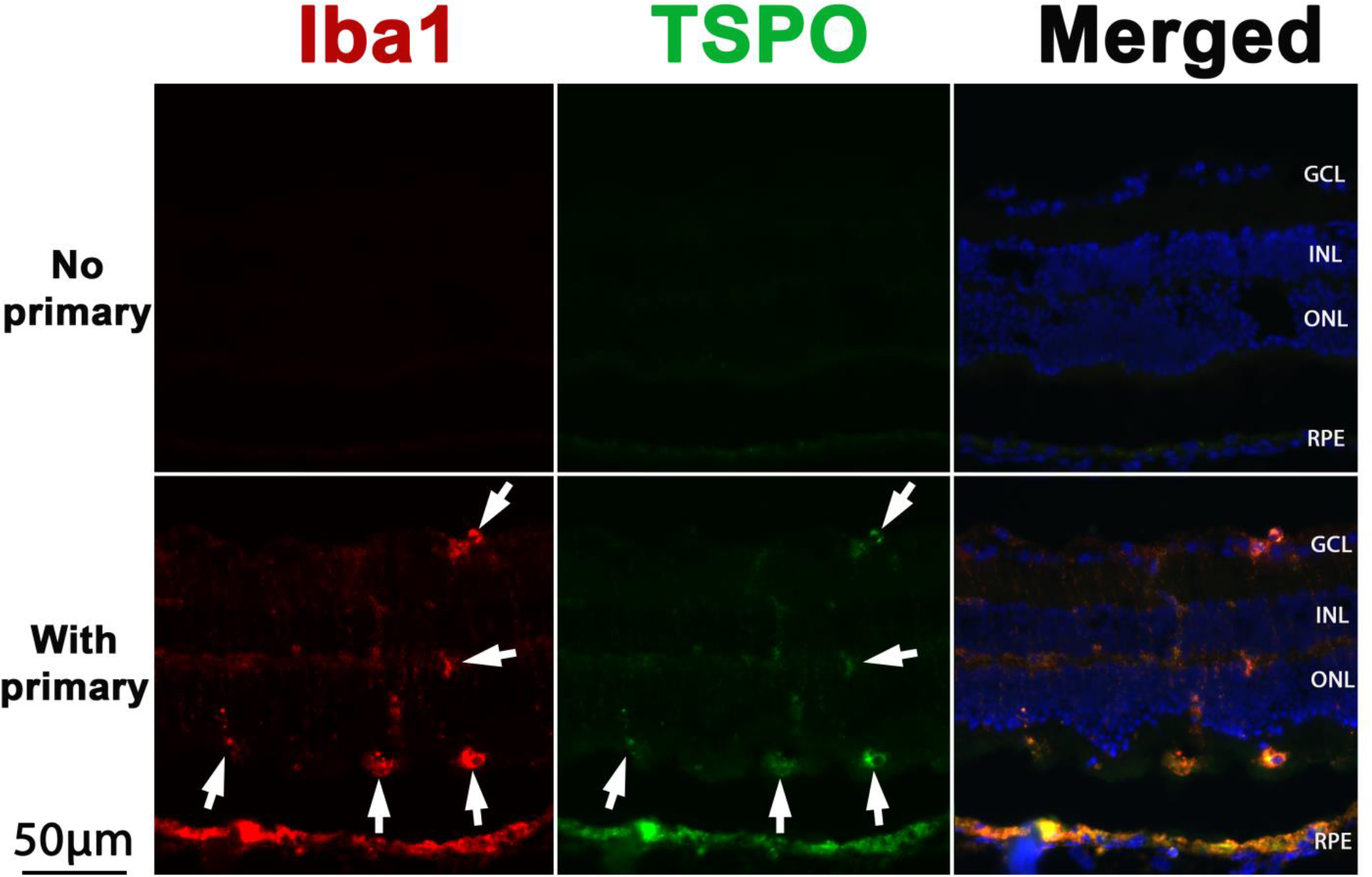
 Figure 2 of
Song, Mol Vis 2017; 23:210-218.
Figure 2 of
Song, Mol Vis 2017; 23:210-218.