Figure 4. Fundus autofluorescence (FAF) photographs and spectral-domain optical coherence tomography (OCT) scans of patients with GUCA1A mutations. Family MTP434 FAF, mild perifoveal hyperautofluorescence in IV:3; small, round, hypoautofluorescent lesion in
IV:1; and no autofluorescence in the macular lesion in III:2. Family MTP434 OCT, complete (in IV:1) or near-complete (in III:2)
foveal atrophy; a thinned ellipsoid zone remained present in IV:3. Family MTP1560 FAF, moderate perifoveal autofluorescence
in IV:1, fovea moderately hypoautofluorescent in III:2 and II:1, and normal autofluorescence in the peripheral retina for
the three patients. Family MTP1560 OCT, thickened ellipsoid zone in the fovea in IV:1, absent ellipsoid zone with thinning
of the outer nuclear layer in the fovea in III:2, and partial foveal atrophy with a hyporeflective zone in both eyes in II:1.
OD, oculus dexter (right eye); OS, oculus sinister (left eye).
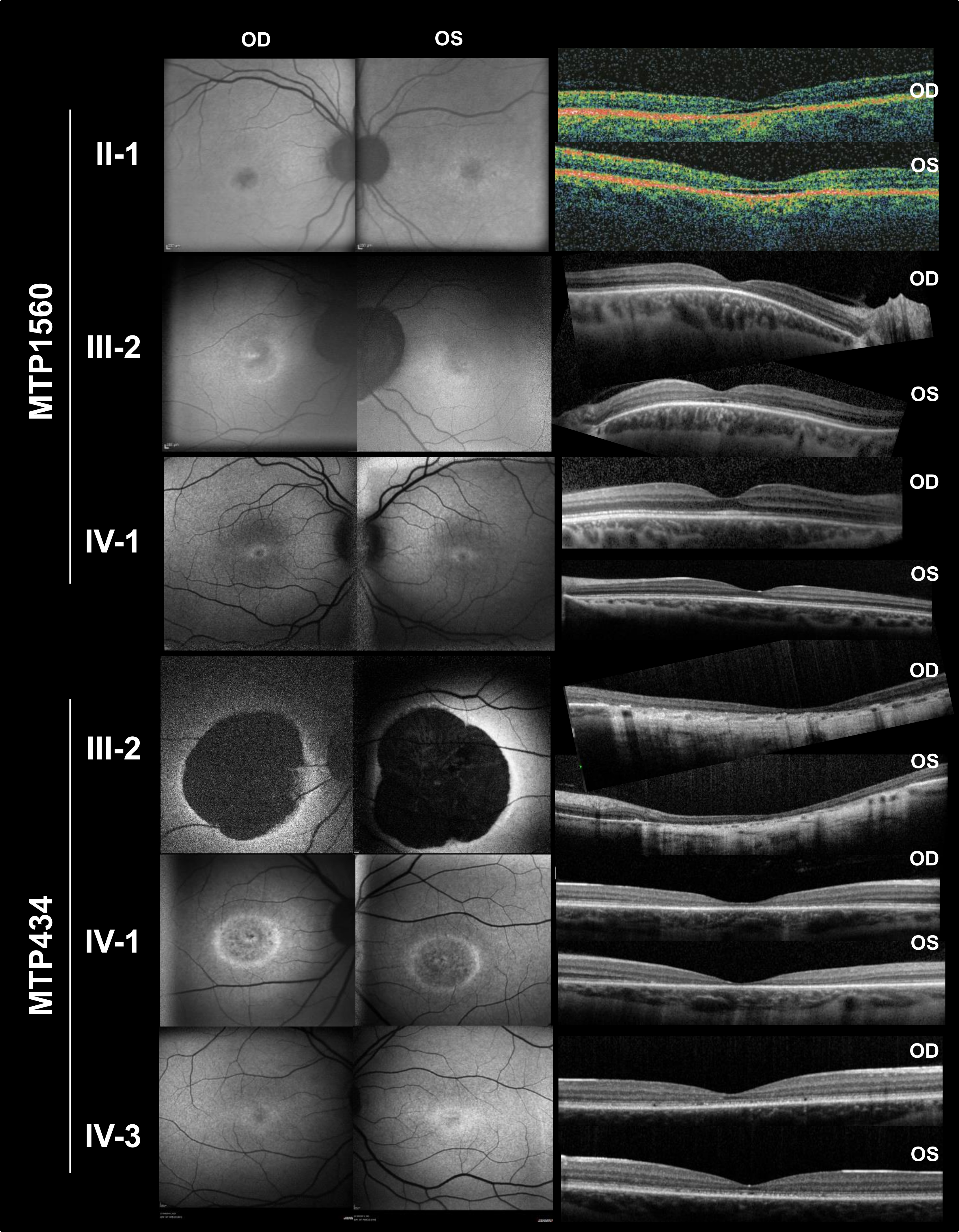
 Figure 4 of
Manes, Mol Vis 2017; 23:198-209.
Figure 4 of
Manes, Mol Vis 2017; 23:198-209.