Figure 2. Summary of the referenced and the novel mutations on the GCAP1 primary structure. Schematic representation of the guanylate
cyclase-activating protein 1 (GCAP1) showing the location of the two novel mutations presented in this study (in bold and
framed) and the known mutations (in bold and underlined; in bold and double underlined when two different mutations are localized
on the same amino acid; in dashed underline for unlikely mutations). The EF-hand domains consist of an alpha-helix (E), a
12-amino-acid loop, and a second alpha-helix (F). The Ca2+ ion is bound in the EF2–4 loops at specific sites (arrowhead); EF1 does not bind calcium. The mutations associated with a
macular dystrophy (MD) phenotype are displayed with a hashtag. EF1–4, EF-hand domains; Myr, N-terminal myristoylation.
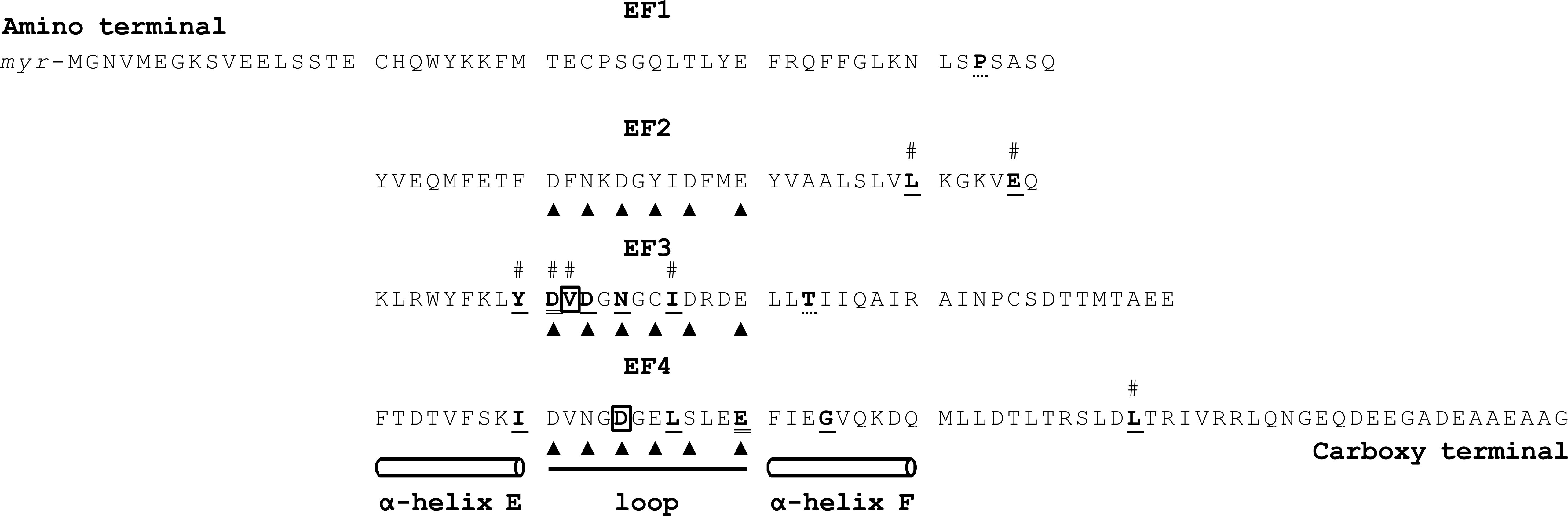
 Figure 2 of
Manes, Mol Vis 2017; 23:198-209.
Figure 2 of
Manes, Mol Vis 2017; 23:198-209.