Figure 3. Zonula occludens-1 (ZO-1) distribution. A: In wild-type (WT) samples, ZO-1 localized to the epithelial–fiber interface and to the vertices of the most peripheral fibers.
B: ZO-1 staining was enriched on the long and short sides of inner cortical fibers (about 150–200 μm from the surface) with
a connexin (Cx)-like staining pattern. C: ZO-1 appeared on the epithelial–fiber interface and the vertices of knock-in (Ki) peripheral fibers. D: ZO-1 was not detected in inner cortical fibers of Ki lenses. Scale bars: 5 μm.
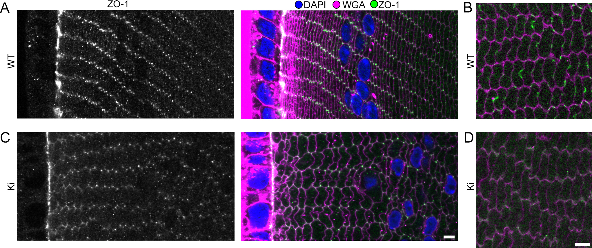
 Figure 3 of
Wang, Mol Vis 2017; 23:160-170.
Figure 3 of
Wang, Mol Vis 2017; 23:160-170.