Figure 3. Clinical investigations of the patient with a homozygous mutation in TTLL5. A: Fundus photographs of the right (left side) and left (right side). There is no macular atrophy or pigment deposits. B, C: The infrared reflectance images (B) and fundus autofluorescence (C) show a moderate increase in perifoveal autofluorescence. D: Optical coherence tomography of the right (up) and left (down) eyes with a selection of peripheral (left) and macular (right)
slices for each side show that the periphery is normal while there are subtle changes in the perifoveal area with attenuation
of the ellipsoid zone. E: The kinetic (peripheral isopter) and static visual field shows a normal peripheral visual field with a general decrease
in retinal sensitivity in the central 30° in accordance with cone dysfunction. F: The International Society for Clinical Electrophysiology of Vision (ISCEV) electroretinogram (ERG) indicates a normal rod
function (dark-adapted ERG) while the amplitude values of the cone function (light-adapted ERG) corresponded to 30% of the
normal values.
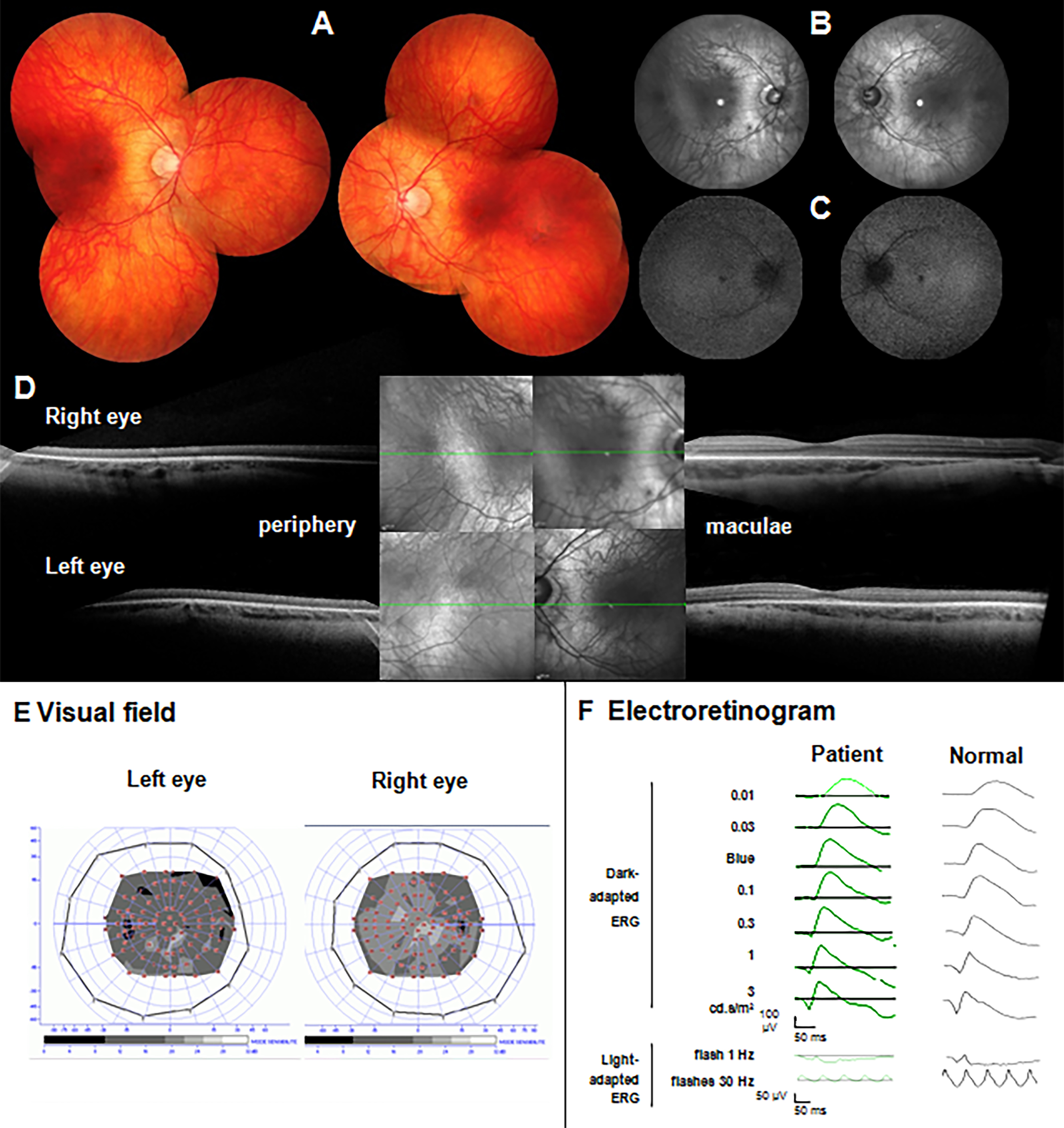
 Figure 3 of
Dias, Mol Vis 2017; 23:131-139.
Figure 3 of
Dias, Mol Vis 2017; 23:131-139.