Figure 7. Hyperosmotic expression of NFAT5 protein in RPE cells. A: The level of NFAT5 protein (170 kDa) was determined by western blot analysis of cytosolic extracts of cells which were cultured
6 h in iso- (control; Co) and hyperosmotic (+100 mM NaCl) media in the absence and presence of the following inhibitory agents:
the phospholipase A2 (PLA2) inhibitor 4-bromophenacyl bromide (Bromo; 30 µM), the phospholipase Cγ (PLCγ) inhibitor U73122 (4 µM), the P2Y1 receptor antagonist MRS2179 (30 µM), and the nuclear factor κB (NF-κB) inhibitor caffeic acid phenethyl ester (CAPE; 5 µM).
Equal amounts of total protein (35 µg) were used for separation. β-Actin (45 kDa) was used as control for equal protein loading.
B: Cellular level of NFAT5 protein, as determined by densitometrical analysis of western blots. Vehicle control was made with
dimethyl sulfoxide (DMSO; 0.1%). Means ± standard error of the mean (SEM) of 3–7 independent experiments using cell lines
from different donors. Significant difference versus unstimulated control: *p<0.05. Significant difference versus NaCl control: ●p<0.05.
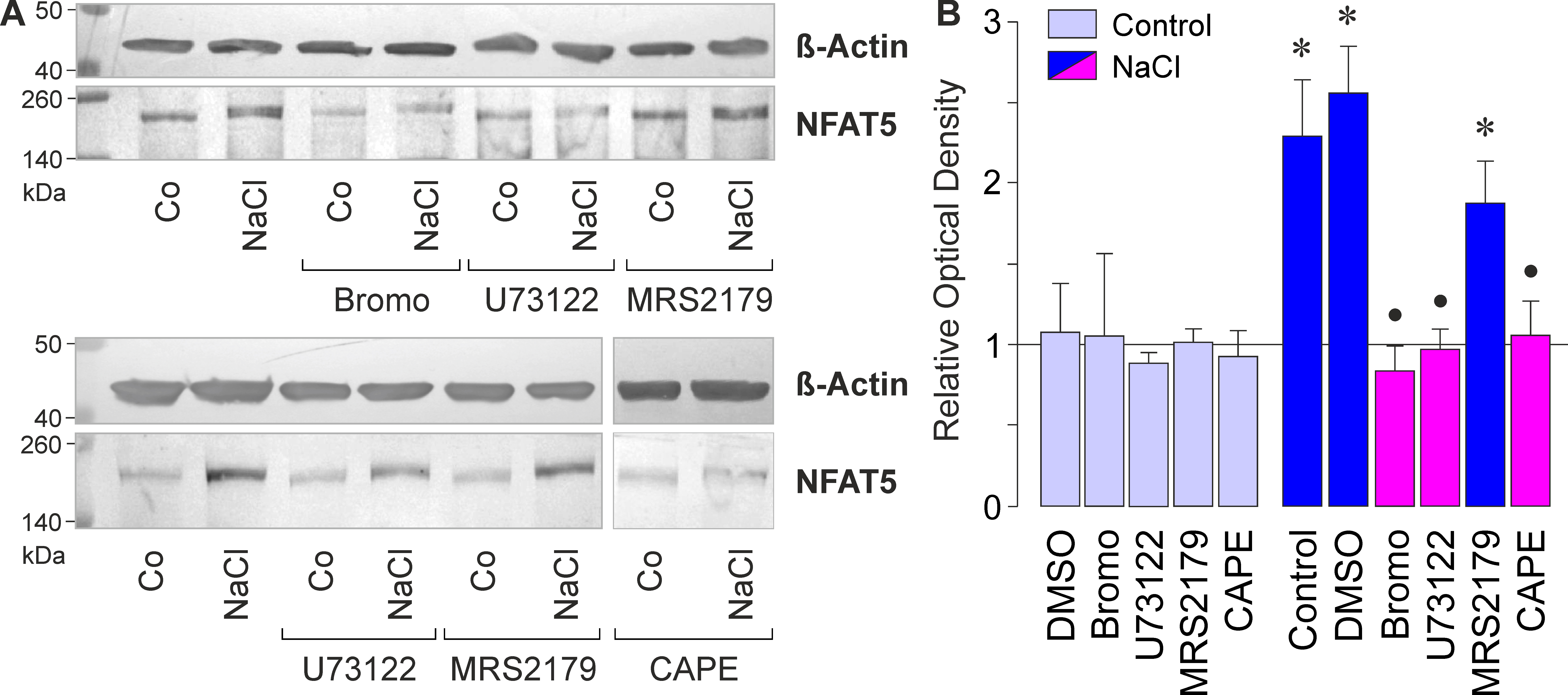
 Figure 7 of
Hollborn, Mol Vis 2017; 23:116-130.
Figure 7 of
Hollborn, Mol Vis 2017; 23:116-130.