Figure 6. Extracellular hyperosmolarity increases the NFAT5 immunoreactivity in RPE cells. Cultured RPE cells were immunolabeled with
antibodies against NFAT5 (green) and vimentin (red). Cell nuclei were stained with Hoechst 33,342 (blue). A: Cells were cultured 6 h in iso- (control; left) and hyperosmotic (+100 mM NaCl) medium (right), respectively. Note the increased NFAT5 immunoreactivity and the altered shape of cells cultured in hyperosmotic medium
compared to control cells. B: In the presence of the phospholipase Cγ (PLCγ) inhibitor U73122 (4 µM), hyperosmotic medium did not cause an increase of
NFAT5 immunoreactivity compared to control. C: Negative control obtained by omitting of the primary antibodies. The cells were cultured 6 h in hyperosmotic (+100 mM NaCl)
medium. No unspecific labeling was observed following cell culture in iso-osmotic medium (not shown). Bars, 20 µm.
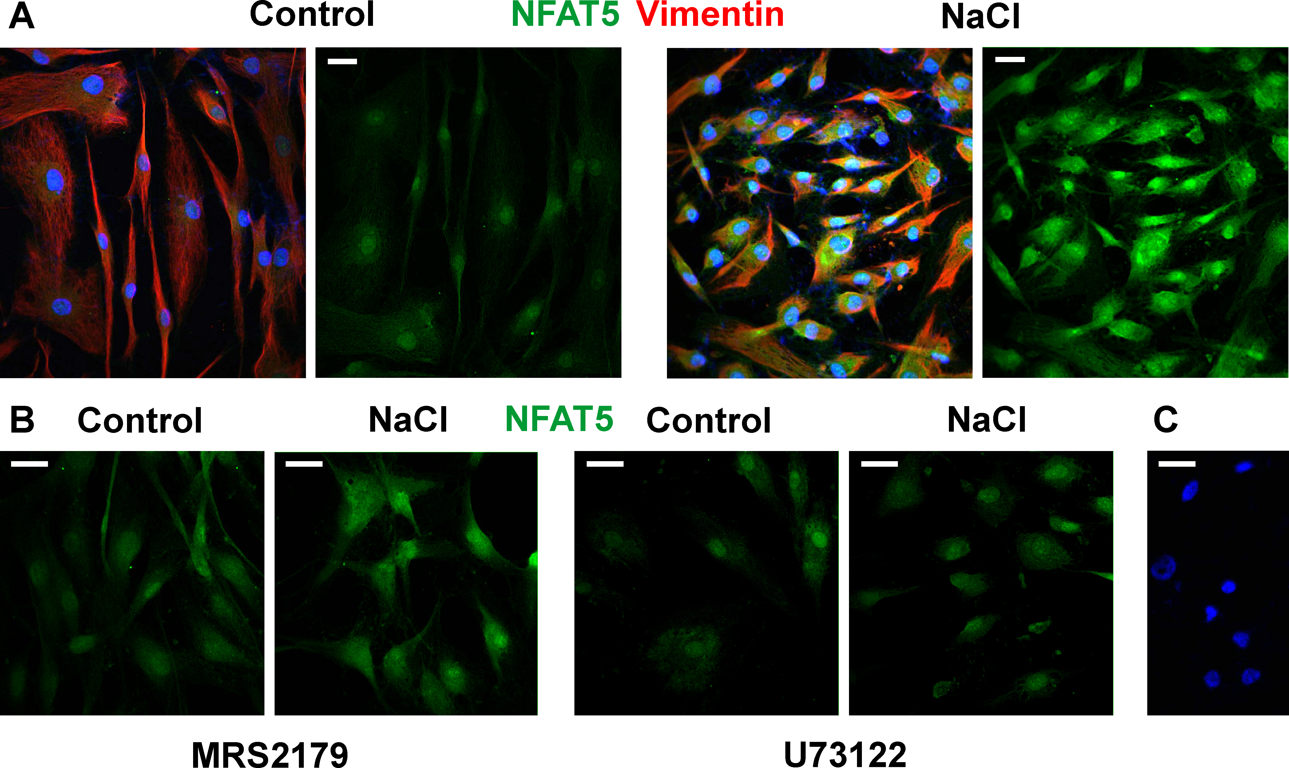
 Figure 6 of
Hollborn, Mol Vis 2017; 23:116-130.
Figure 6 of
Hollborn, Mol Vis 2017; 23:116-130.