Figure 7. Low physiologic oxygen conditions increased the mRNA and protein expression of lactate transporter MCT4 in choroidal melanocytes.
A: Heatmap representation of monocarboxylate transporter (MCT4) expressed by choroidal melanocytes grown at 21% or 3% O2 (n = 3/group). Data are also presented for the housekeeping mRNA golgin A1 (GOLGA1), C-terminal binding protein 1 (CTBP1),
and beta-2-microglobulin (B2M) (upper panel). Transcripts indicated in dark blue correspond to those whose expression is low,
whereas the highly expressed transcripts are shown in orange and red. Corresponding linear signals obtained for the MCT4 mRNA
at 21% (black) or 3% (gray) O2 and normalized with the housekeeping mRNA are displayed as the ratio of the linear signals using a column bar graph (lower
panel; mean ± standard error of the mean [SEM]). *p<0.05, Student t test. B: Western blotting conducted on protein extracts from choroidal melanocytes (M1–M3) or uveal melanoma (UM) cells (T111, T131,
T132) expanded at 21% (odd numbers) or 3% (even numbers) O2 (n = 3/group) using an antibody against MCT4 (upper panel; 43 kDa). The relative amount of total MCT4 protein per lane was
calculated at 21% (black) or 3% (gray) O2 and then normalized with the Stain-Free signal intensities. Data are presented as relative expression of MCT4 using a column
bar graph (lower panel; mean ± SEM). *p<0.05, ** p<0.01, ***p<0.001, Student t test. ND, no detectable bands.
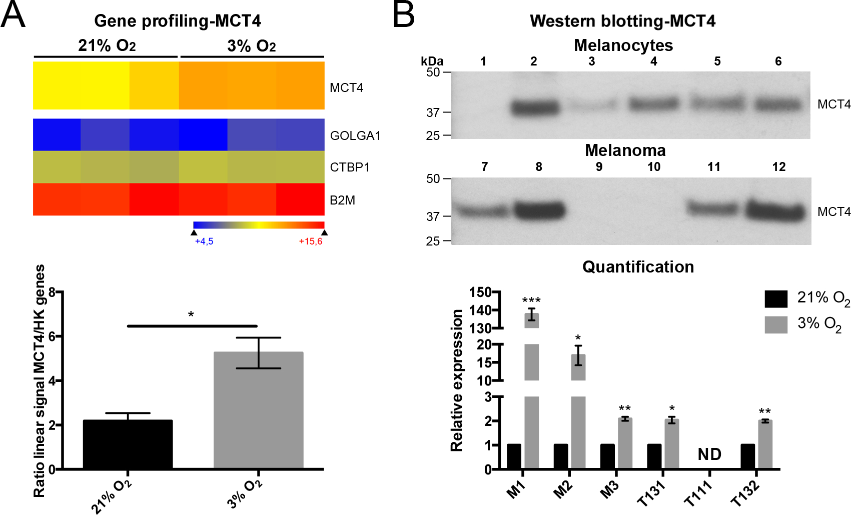
 Figure 7 of
Weidmann, Mol Vis 2017; 23:103-115.
Figure 7 of
Weidmann, Mol Vis 2017; 23:103-115.