Figure 4. Low physiologic oxygen conditions shorten the doubling time of choroidal melanocytes and increase their proliferative capacity.
A: The doubling time of the choroidal melanocytes (n = 3) expanded under 21% (•) or 3% (■) O2 conditions was determined at three consecutive passages (mean ± standard error of the mean [SEM]). *p<0.05, **p<0.005, the
Student t test. B: The percentage of choroidal melanocytes (n = 3) in the active phases of the cell cycle was determined with immunofluorescence
analyses using Ki67 on cells exposed to 21% (black) or 3% (gray) O2. The Ki67-positive cells were counted, and the total number of 4',6-diamidino-2-phenylindole (DAPI)-stained nuclei was used
for normalization. **p<0.005, ***p<0.0005, Mann–Whitney test. C: The doubling time of uveal melanoma (UM) cells (n = 3) cultured under 21% (•) or 3% (■) O2 conditions was determined at three consecutive passages (mean ± SEM). *p<0.05, ***p<0.0005, the Student t test. D: The percentage of UM cells (n = 3) in the mitotic phase of the cell cycle was determined with immunofluorescence analyses
using phosphohistone H3 (PH3) on cells exposed to 21% (black) or 3% (gray) O2. The PH3-positive cells were counted, and the total number of DAPI-stained nuclei was used for normalization.
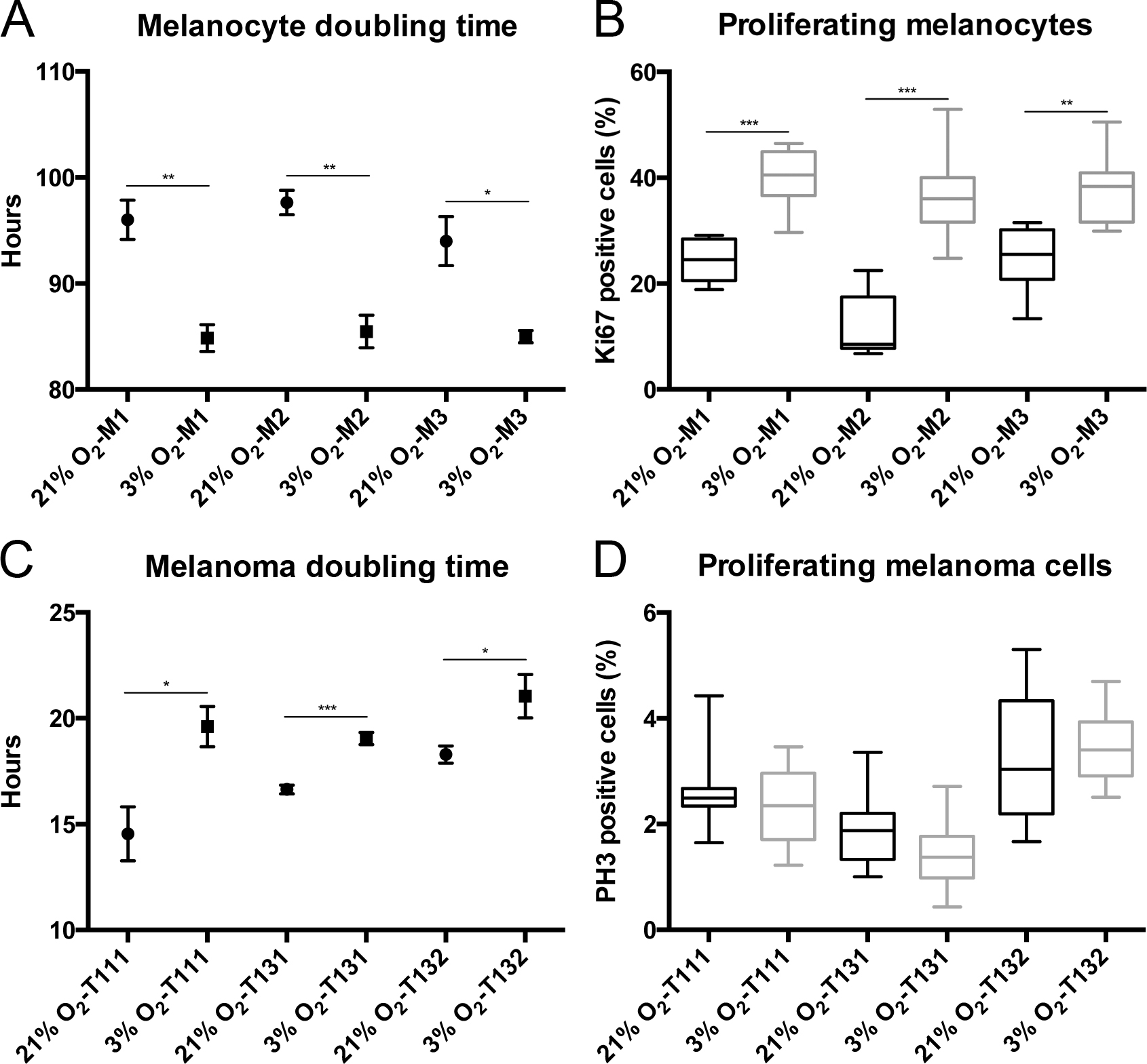
 Figure 4 of
Weidmann, Mol Vis 2017; 23:103-115.
Figure 4 of
Weidmann, Mol Vis 2017; 23:103-115.