Figure 3. Low physiologic oxygen conditions do not increase cell senescence. A: Western blotting was conducted on protein extracts from choroidal melanocytes (n = 3) expanded at 21% (wells 1, 3, 5) or
3% (wells 2, 4, 6) O2 using an antibody against caveolin-1 (CAV1; senescence marker, upper panel; 21 kDa). The relative amount of total CAV1 proteins
per lane was calculated at 21% (black) or 3% O2 (gray) and then normalized with the Stain-Free signal intensities. Data are presented as the expression of CAV1 using a column
bar graph (lower panel; mean ± standard error of the mean [SEM]). B: Western blotting was conducted on protein extracts from uveal melanoma (UM) cells (n = 3) expanded at 21% (wells 1, 3, 5)
or 3% (wells 2, 4, 6) O2 using an antibody against CAV1 (upper panel). The relative amount of total CAV1 proteins per lane was calculated at 21% (black)
or 3% O2 (gray) and then normalized with the Stain-Free signal intensities. Data are presented as the expression of CAV1 using a column
bar graph (lower panel; mean ± SEM).
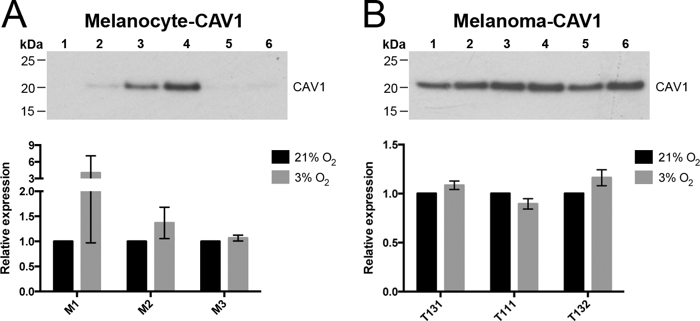
 Figure 3 of
Weidmann, Mol Vis 2017; 23:103-115.
Figure 3 of
Weidmann, Mol Vis 2017; 23:103-115.