Figure 6. HLEC viability assay with or without Aβ intervention under oxidative stress. A: Inverted microscope investigation of cell morphology with or without β-amyloid (Aβ) 1–40 pretreatment under oxidative stress.
B: Human lens epithelial cell (HLEC) viability assay with or without Aβ 1–42 pretreatment under oxidative stress. C: HLEC viability assay with or without Aβ 1–40 pretreatment under oxidative stress. The concentration of H2O2 was 200 μM. Aβ concentrations were 0.2 nM to 10 nM as listed in (B) and (C). The data are the means ± standard deviation (SD). In each group, three independent tests were performed. *p<0.05, **p<0.01,
***p<0.001 comparing the Aβ intervention groups with the vehicle control group, with one-way ANOVA and least significant difference
(LSD) tests.
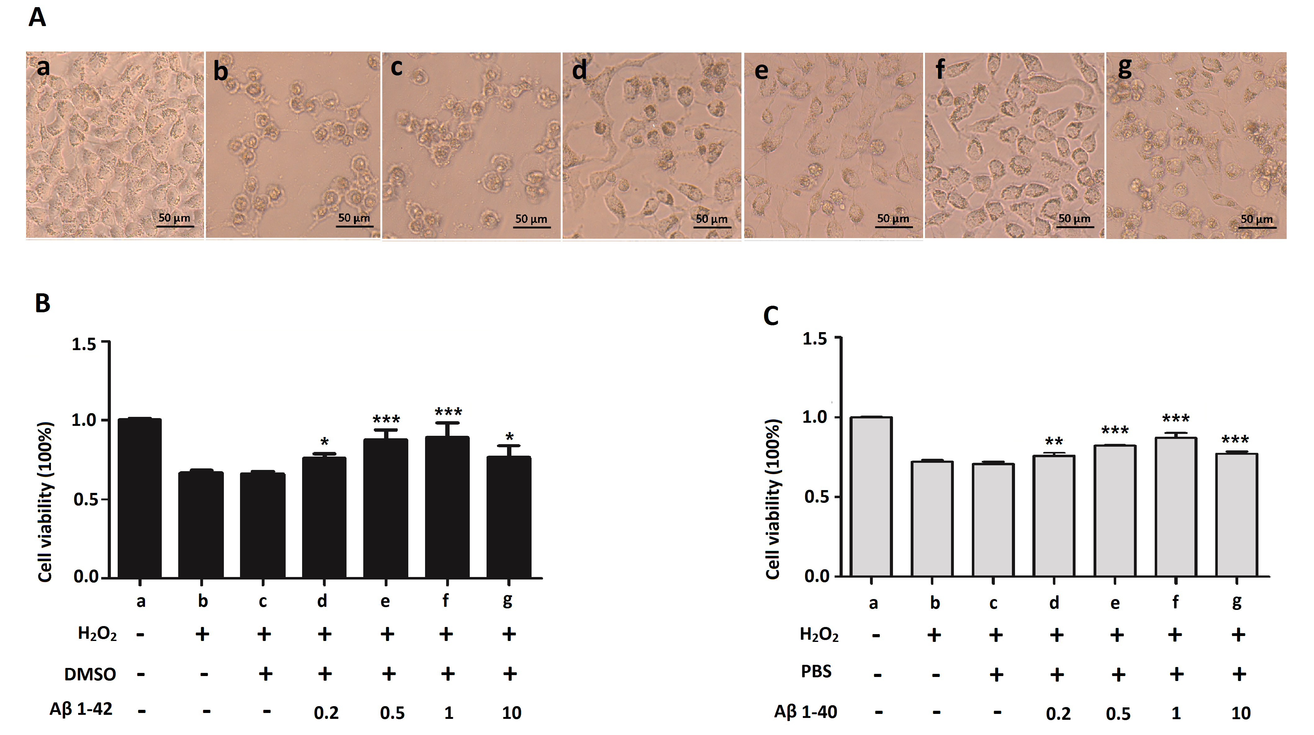
 Figure 6 of
Xu, Mol Vis 2017; 23:1015-1028.
Figure 6 of
Xu, Mol Vis 2017; 23:1015-1028.