Figure 2. Dynamic changes in ADORs affected by 7-MX at the mRNA level. Human RPE cells were treated with 10 μmol/l 7-methylxanthine
(7-MX) for 24, 48, and 72 h. The mRNA levels of ADORA1 (A), ADORA2A (B), and ADORA2B (C) was detected with quantitative PCR (qPCR). GAPDH was used as the internal control. n=3. * p<0.05, ** p<0.01, and ***p<0.001
compared with 0 h; ## p<0.01 compared with 24 h; ▲ p<0.05 and ▲▲ p<0.01 compared with 48 h.
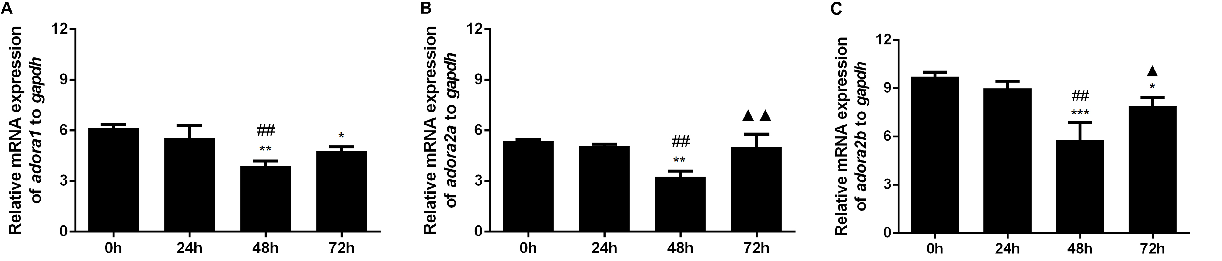
 Figure 2 of
Wan, Mol Vis 2017; 23:1006-1014.
Figure 2 of
Wan, Mol Vis 2017; 23:1006-1014.