Figure 2. Epac1 can be knocked out in vascular endothelial cells. A: The agarose gel image from the mouse ear punch samples shows the effective knockout of exchange protein for cAMP 1 (Epac1)
by Cdh5 Cre. Expected band sizes: Epac mutant 457 bp, wild-type 276 bp, Cdh5 Cre 300 bp. B: A significant reduction in the Epac1 protein levels from the whole retinal lysates. n = 5 mice in each group. C: Imaging data show the cellular localization of Epac1 in the retina from the Epac1 floxed and Epac1 Cre-Lox mice. The top
image is from the Epac1 floxed mice, and the bottom image is from the Epac1 Cre-LoxP mice. On the right is a larger image
showing the overlap of Epac1 and B4 staining only in the floxed mice. Epac1 staining is in red, vascular staining (isolectin
B4) is in green, and cell nuclei is in blue. Scale bar = 50 μm for left images; scale bar = 20 μm for enlarged images.
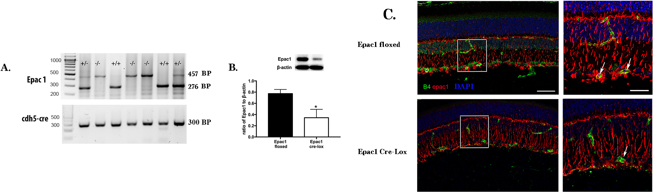
 Figure 2 of
Liu, Mol Vis 2017; 23:1-7.
Figure 2 of
Liu, Mol Vis 2017; 23:1-7.