Figure 3. Migration trajectories of corneal epithelial cells in reconstructed corneal cultures. A: Migration index of non-random migration of corneal epithelial cells plated onto corneal basement membranes that had been
de-epithelialized by scraping. The migration index is obtained by dividing the mean angle of cell migration by the radial
direction to the corneal center under the experimental conditions described by the mean angle of cell migration of control
cells on a tissue culture dish. Therefore, a migration index of 1 represents random migration; a migration index of 0 represents
perfect radial migration toward the center of the cornea. Abbreviations: ns, nonsignificant. * represents p<0.05; ** represents
p<0.01. All cells made migration errors, but wild-type mouse epithelial cells showed non-random migration biased radially
when plated onto wild-type corneas. Conversely, Pax6+/− corneal epithelial cells plated onto wild-type stroma and wild-type cells treated with RP cAMPS to inhibit cAMP-mediated
intracellular signaling migrated randomly. B: Migration tracks of wild-type mouse primary corneal epithelial cells on de-epithelialized wild-type corneas. Composite from
three overlaid corneas from three separate experiments (pink, red, blue). For each cornea. the point of most likely convergence
(i.e., with minimum mean migration error across all cells migrating on that cornea) is indicated (solid circles). C: Migration tracks of wild-type primary mouse corneal epithelial cells on de-epithelialized wild-type corneas with 40 μM RP
cAMPS added to the culture medium. Composite from three overlaid corneas from three separate experiments (pink, red, blue).
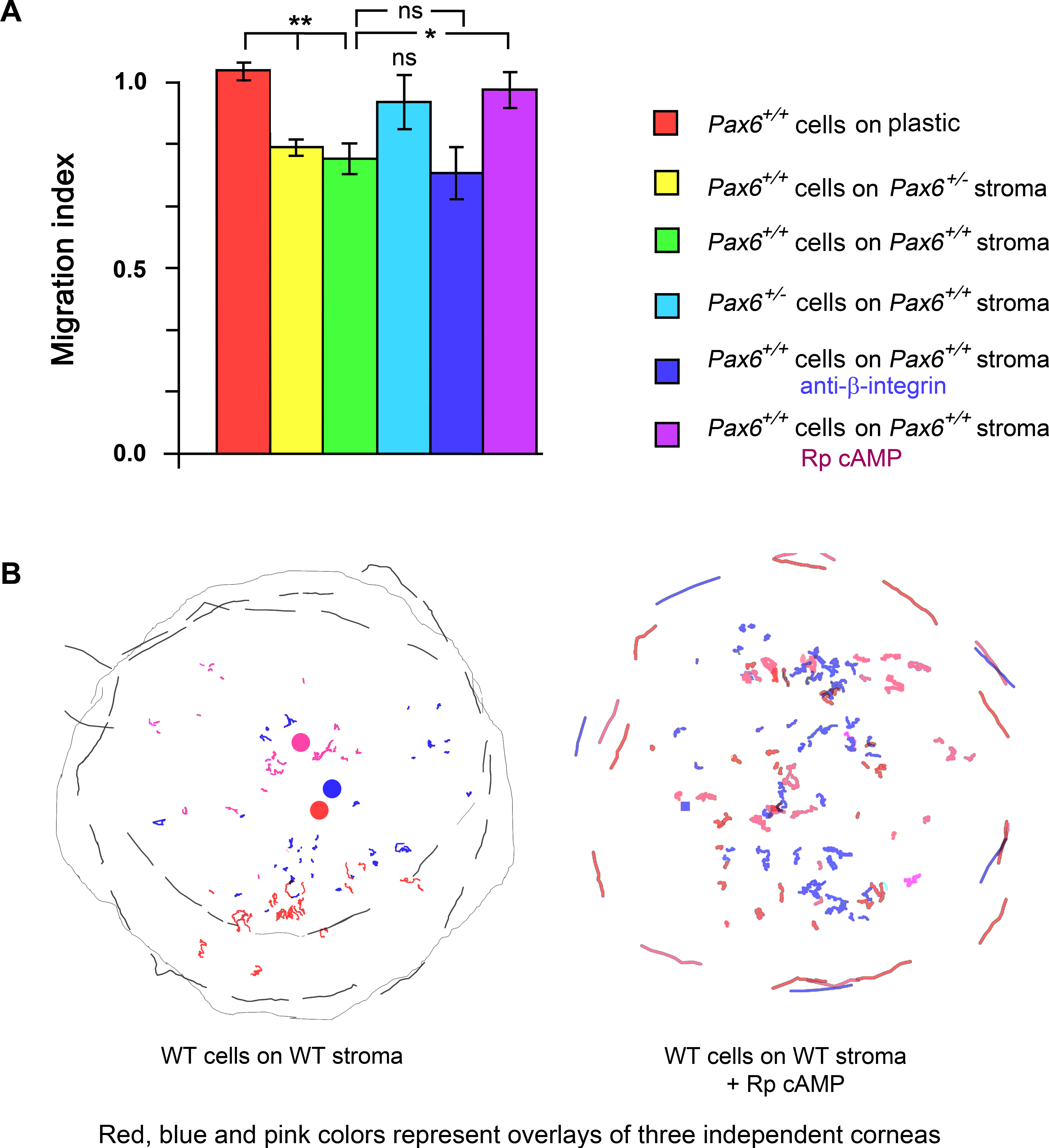
 Figure 3 of
Walczysko, Mol Vis 2016; 22:990-1004.
Figure 3 of
Walczysko, Mol Vis 2016; 22:990-1004.