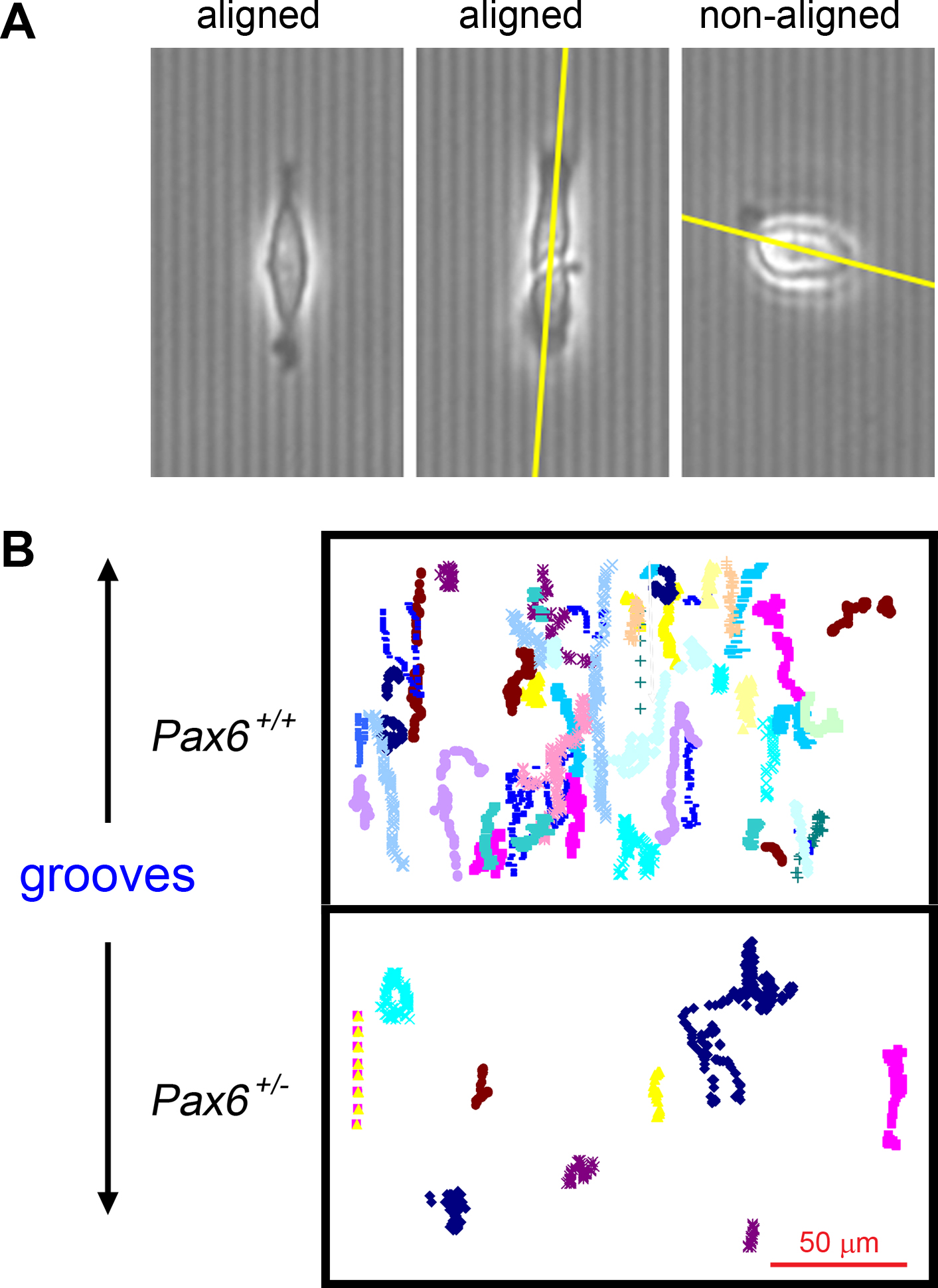
Figure 2. Alignment and migration of mouse corneal epithelial cells on grooved quartz substrata.
A: Images of aligned (left and middle) and non-aligned (right) primary mouse corneal epithelial cells on 2 μm grooved quartz
surfaces. Yellow lines indicate the long axis of the cells. See
Table 1 for quantification.
B: Tracks of cells migrating on 2 μm quartz grooves. Scale bar represents 50 μm.
 Figure 2 of
Walczysko, Mol Vis 2016; 22:990-1004.
Figure 2 of
Walczysko, Mol Vis 2016; 22:990-1004.