Figure 1. Corneal epithelial wound healing in vivo in mice. A: Images of wild-type (left) and Pax6+/− (right) adult mouse eyes shortly after corneal epithelial wounding. The wound areas of the approximately 1 mm diameter wounds
have been highlighted for visualization. The boundary of the 1 mm diameter circular epithelial wound is visible. Vascularization
of the cornea is a variable feature of the chronic wound healing phenotype of Pax6+/− mice (discussed in the text). B: Rates of epithelial wound healing in wild-type (left) and Pax6+/− (right) adult mice. Mean rates of migration are given over 0–2 h, 0–6 h, 0–12 h, and 0–21 h. Means ± standard deviation are
shown. Number of wounds are 0–2 h, Pax6+/+ = 10, Pax6+/− = 8; 0–6 h, Pax6+/+ = 2, Pax6+/− = 2; 0–12 h, Pax6+/+ = 8, Pax6+/− = 6; and 0–21 h, Pax6+/+ = 12, Pax6+/− = 12. The wound healing profiles, with an initial retraction followed by re-epithelialization, are similar, but the migration
of the Pax6+/− corneal epithelial cells is statistically significantly slower than in the wild-type in the latter healing phases.
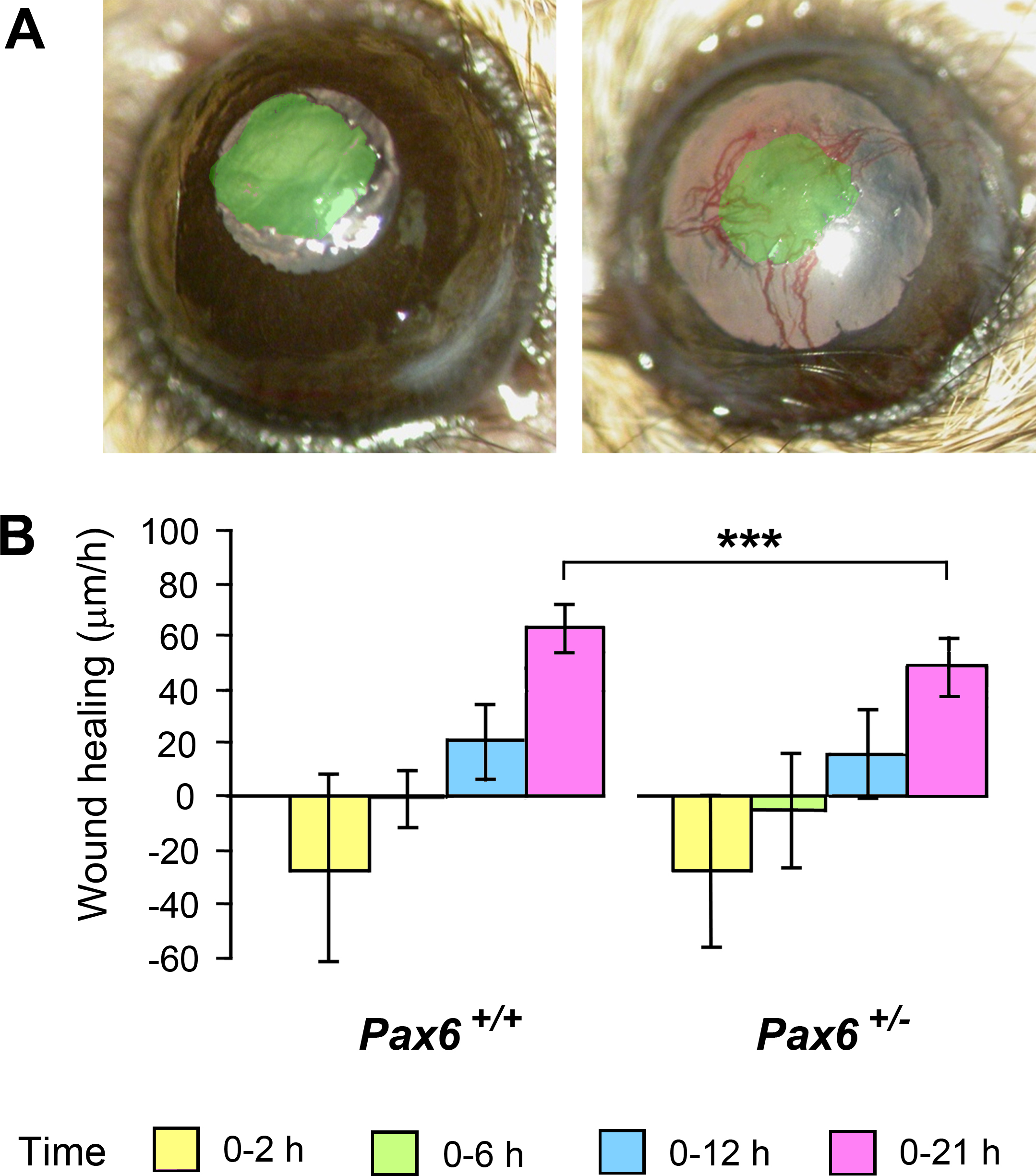
 Figure 1 of
Walczysko, Mol Vis 2016; 22:990-1004.
Figure 1 of
Walczysko, Mol Vis 2016; 22:990-1004.