Figure 7. Colocalization of vimentin and CP49. A montage of four pairs of images taken from a lens epithelial explant double-labeled
with antibodies to CP49 (A–D), and vimentin (A’–D’). The vermiform structure is clearly labeled with antibodies to CP49 in the upper panel, while the lower panel shows that
vimentin is absent (or at much lower levels) in those same structures (marked by red arrows; scale bar = 10 μm).
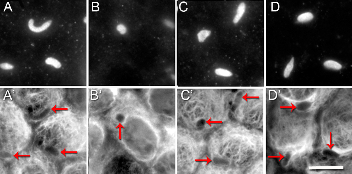
 Figure 7 of
FitzGerald, Mol Vis 2016; 22:970-989.
Figure 7 of
FitzGerald, Mol Vis 2016; 22:970-989.