Figure 6. Actin and CP49 colocalize in the vermiform structure. A: Mouse lens epithelial explant labeled with phalloidin identifies filamentous actin (red channel). Arrows identify six examples
(of several dozen) of the vermiform structure. B: The same field of view as in A, but captured in the green channel, which identifies labeling for CP49. The same six examples of the vermiform structure
are demarcated with arrows. C: Phalloidin labeling of a lens epithelial explant from a CP49 knockout mouse shows a rich network of actin filaments (red
channel) but no evidence of the vermiform structure (scale bar = 40 μm).
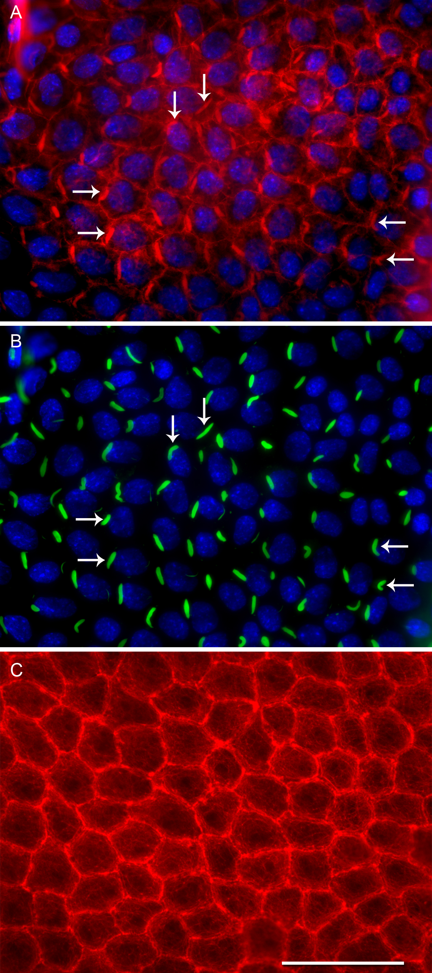
 Figure 6 of
FitzGerald, Mol Vis 2016; 22:970-989.
Figure 6 of
FitzGerald, Mol Vis 2016; 22:970-989.