Figure 4. Electron microscopy. A: An overview of parts of three mouse lens epithelial cells (“N” = nucleus). The capsule is indicated by the white arrow at
top and the CP49-reactive structures by the black arrows (scale bar = 8 μm). B: Higher magnification view of the vermiform structure shows CP49 immunoreactivity, revealed by the presence of 10 nm gold
particles. This structure commonly exhibits an electron lucent region (example, black arrow). The structure is surrounded
by a meshwork of filaments (white arrow). Scale bar = 1 μm. C: Routine transmission electron microscopy (TEM) of the lens epithelial cell (scale bar = 1 μm). The structure is dense enough
that any substructure is not obvious under routine immersion-fixation conditions. D–E: The “ghosted” lens epithelium (D) and fiber cell (E) display the beaded filaments (BFs) more readily when soluble proteins are allowed to diffuse away. E: A single fiber cell that exhibits BFs (white asterisk) and canonical intermediate filaments (IFs; black asterisk) highlighting
the difference between these two classes of IFs. F: An isolated pair of BFs imaged with negative stain TEM, clearly revealing the BF structure (scale bar = 20 nm). The vermiform
structure of the epithelium (white asterisk, D) does not clearly reveal obvious BFs, possibly due to compaction, or to assembly into some other format.
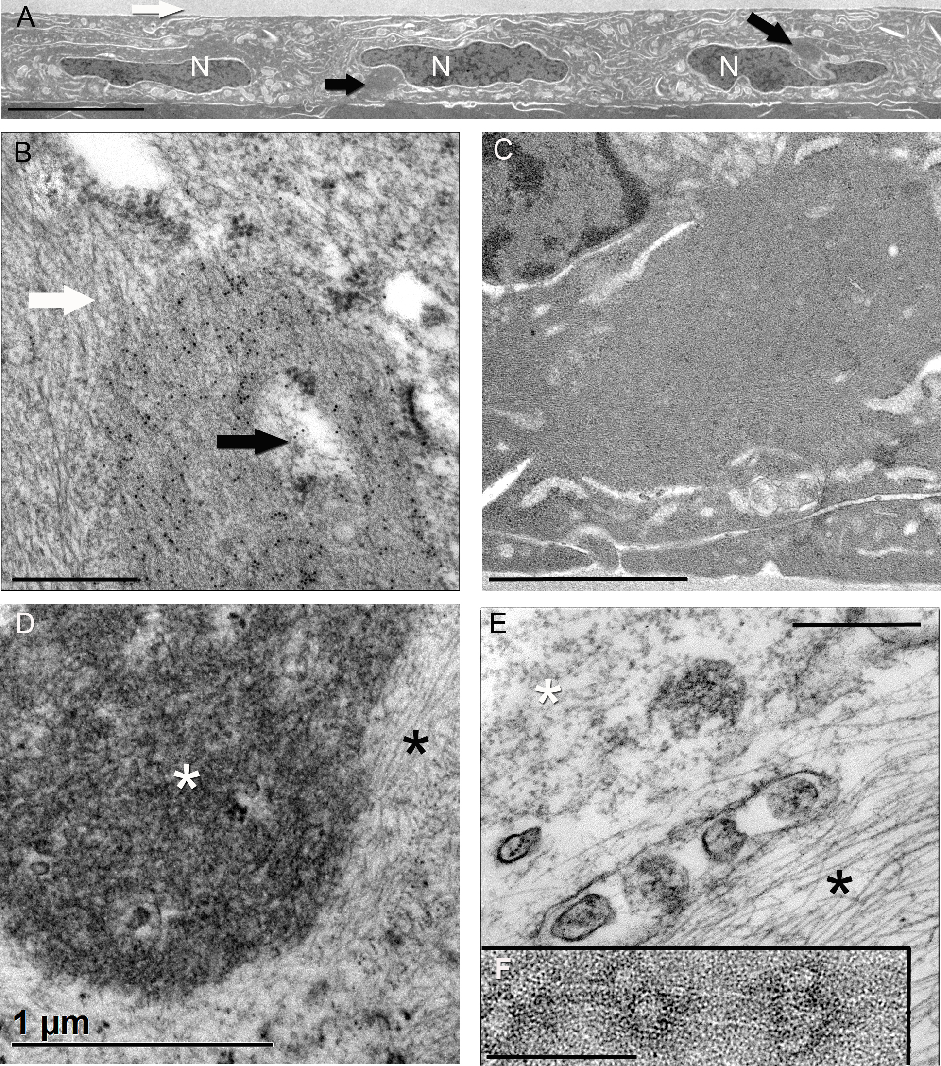
 Figure 4 of
FitzGerald, Mol Vis 2016; 22:970-989.
Figure 4 of
FitzGerald, Mol Vis 2016; 22:970-989.