Figure 3. Spatial distribution of epithelial labeling. A: Paraffin section overview of the anterior lens labeled for CP49 (green channel) and oriented with the anterior pole at the
12 o’clock position. The CP49-reactive epithelial structure becomes smaller and punctate, and then finally disappears near
the coronal equator. The last obvious indication of CP49 reactivity in epithelial cells occurs at about the white arrow (scale
bar = 200 μm). B: Colocalization of EdU (red channel) that labels recently divided cells and the CP49-reactive structure. It is evident that
the loss of the vermiform shape occurs in the region of the lens where the epithelium is mitotically active (scale bar = 150
μm). C: At higher magnification, arrows identify one of the punctate remnants. Deeper in the lens cortex, CP49 reactivity once again
begins to accumulate in the maturing fiber cells (C, right-hand edge) as has been reported previously. Scale bar = 100 μm.
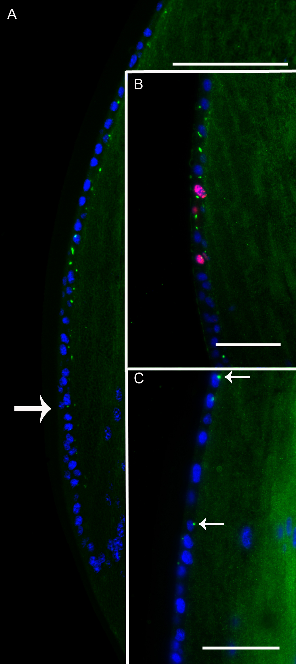
 Figure 3 of
FitzGerald, Mol Vis 2016; 22:970-989.
Figure 3 of
FitzGerald, Mol Vis 2016; 22:970-989.