Figure 1. RSM with integrating cavity absorption meter and actinic LED source. A: Scan phase: The moving slit from the rapid-scanning spectrophotometer (RSM) creates spectral scans at a rate of 100 scans
per second. Light from the RSM is split to each of the cuvette compartments (not illustrated). There, conventional single-pass
rectangular cuvettes are replaced with spherical quartz cuvettes (DeSa Spherical Presentation Chambers, DSPCs). Each DSPC
is surrounded by four light ports. In the present configuration, the incoming light enters through the port opposite the light-emitting
diode (LED). The photomultiplier port is positioned at right angles to both. The spherical cuvette is surrounded by a reflective
material fully scattering the measurement beam as well as the monochromatic light from the LED. The highly reflecting cavity
acts as a multipass cuvette. The effective path length is markedly increased (tens of centimeters) compared to traditional
single-pass cuvettes, and effects of light scatter are eliminated. B: Actinic exposure pulse: A fixed light block creates a dark period within each scan. During this period, the LED is triggered
to flash, and the photomultiplier shutter is temporarily closed. By interleafing scans with LED flashes, light-induced chemical
changes can be followed spectroscopically in real time.
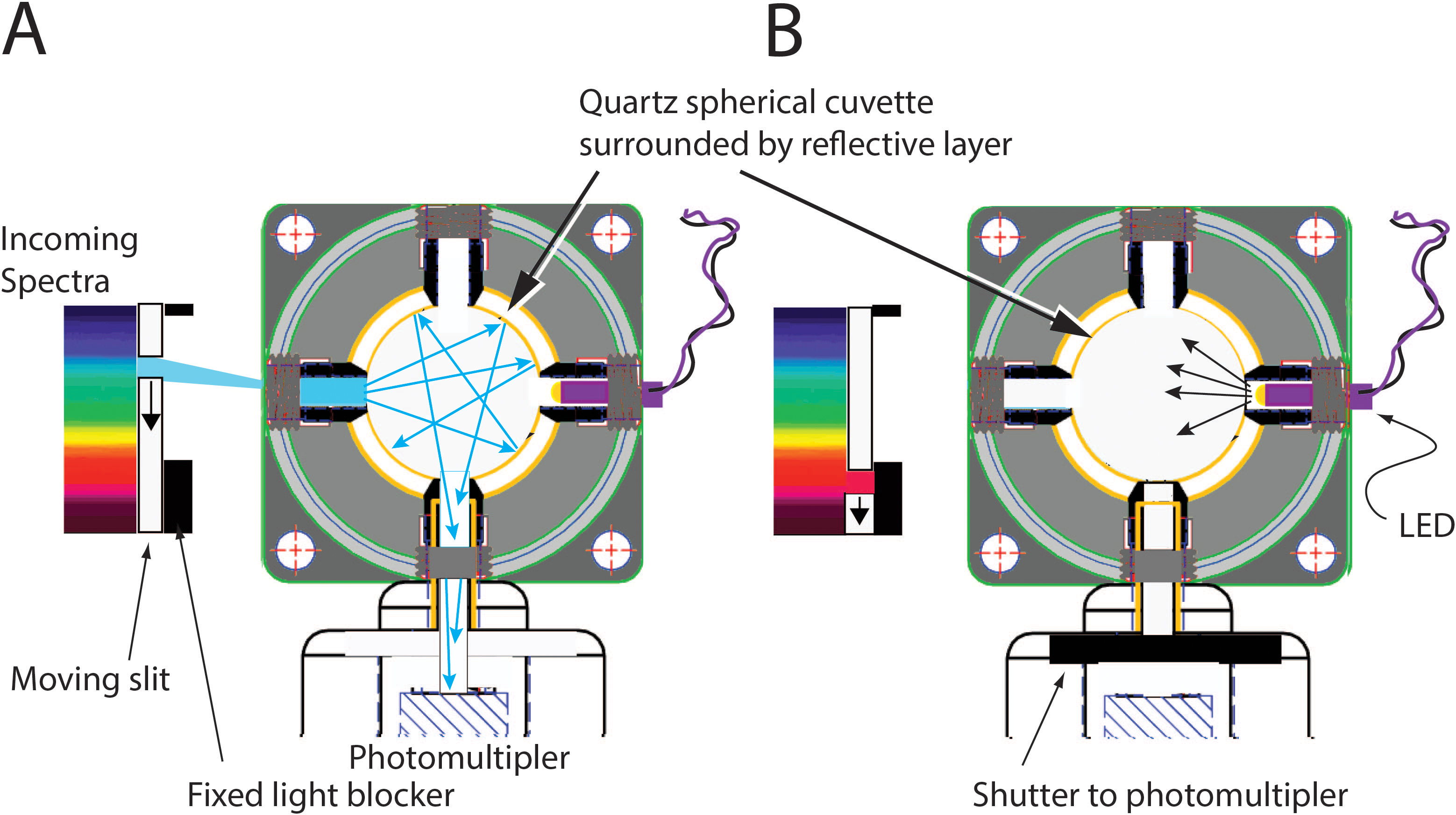
 Figure 1 of
Gonzalez-Fernandez, Mol Vis 2016; 22:953-958.
Figure 1 of
Gonzalez-Fernandez, Mol Vis 2016; 22:953-958.