Figure 3. Images of corneal ulcer with slit-lamp microscopy and NET formation by immunofluorescence. In both specimens from the large
(upper) and small (lower) corneal ulcers, a similar number of neutrophil extracellular traps (NETs) were detected. The arrow
points to the fungal hyphae that exist in immunofluorescence staining. Blue = DNA; red = elastase. Bar: 100 μm.
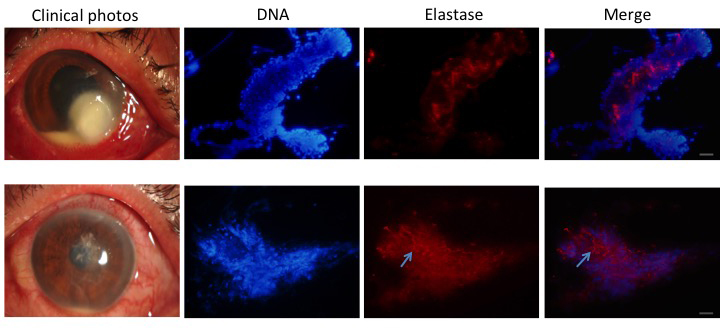
 Figure 3 of
Jin, Mol Vis 2016; 22:944-952.
Figure 3 of
Jin, Mol Vis 2016; 22:944-952.