Figure 1. NETs (arrows) were identified in corneal scrapings of fungal keratitis. DNA (extracellular and nuclear DNA of epithelial cells
and neutrophils) was stained with 4’,6-diamidino-2-phenylindole (blue), and neutrophil elastase was stained red. Bar: 100
μm. The colocalization analysis shows r = 0.412 (Pearson correlation) and r = 0.987 (overlap) by Image J. Arrows point to
the formation of the neutrophil extracellular traps (NETs) in immunofluorescence staining.
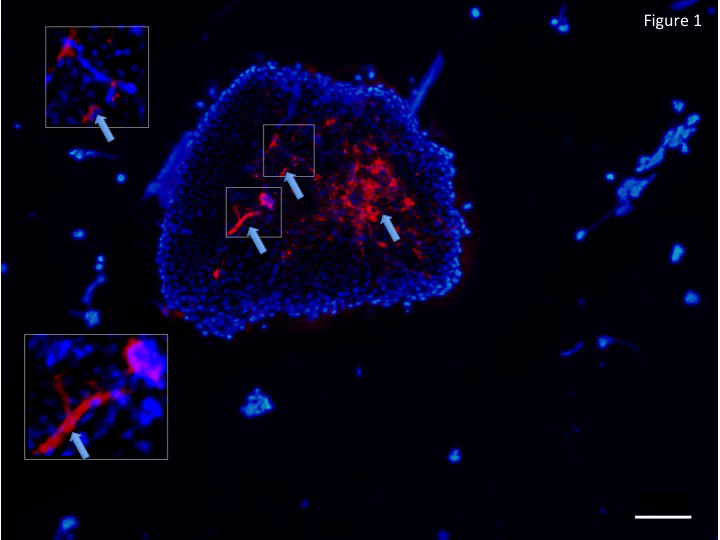
 Figure 1 of
Jin, Mol Vis 2016; 22:944-952.
Figure 1 of
Jin, Mol Vis 2016; 22:944-952.