Figure 5. Fundus photography and fluorescein angiography of the FEVR patients with FZD4 mutations. The patient ID is indicated on the top left of each picture. A: Peripheral avascular zone. B: Falciform retinal fold. C: Peripheral avascular zone and neovascularization. D: Ectopic macular. E: Falciform retinal fold.
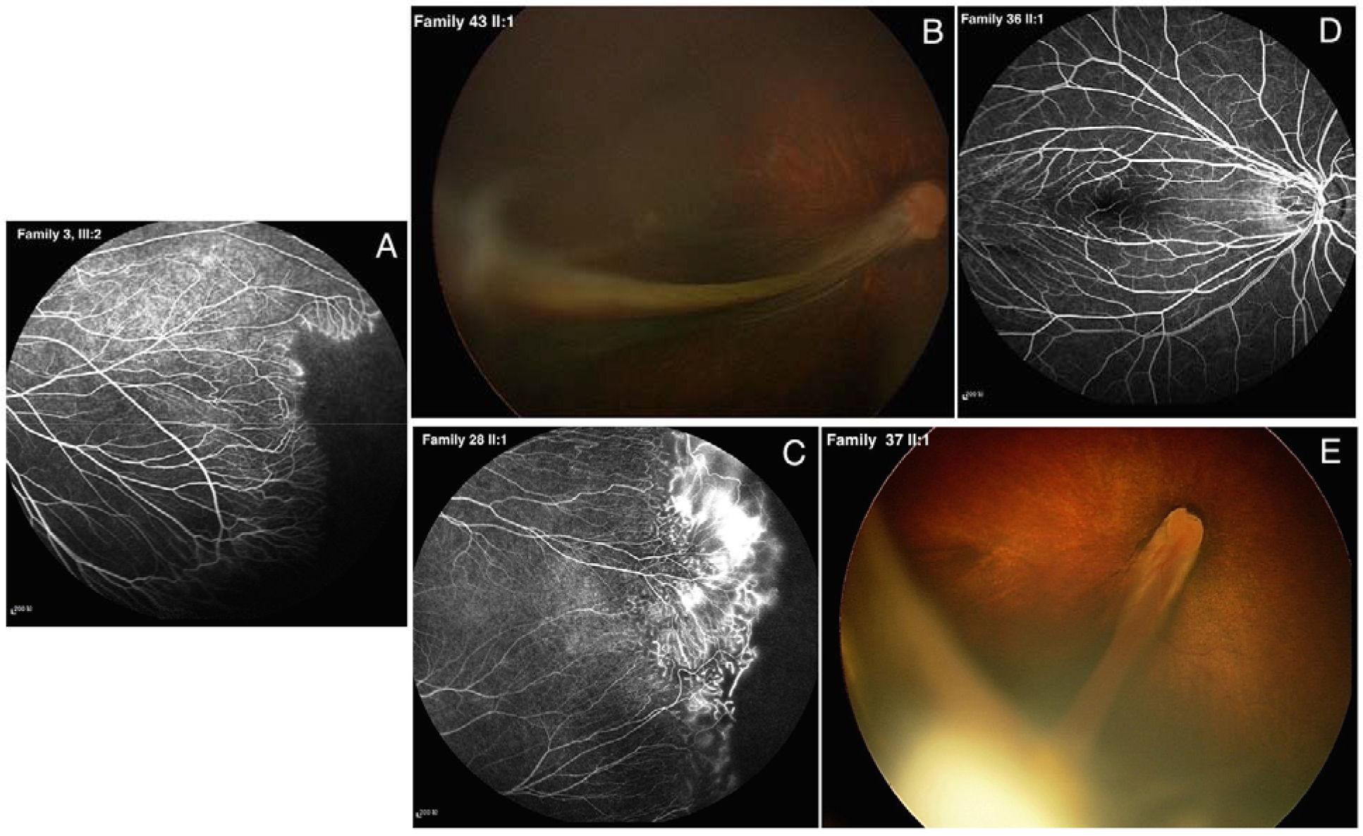
 Figure 5 of
Tang, Mol Vis 2016; 22:917-932.
Figure 5 of
Tang, Mol Vis 2016; 22:917-932.