Figure 1. Description of microdissection methods. Whole eyes were received from the eye bank. A: Orientation of the eye was identified using the superior and inferior oblique muscle insertions and the long posterior ciliary
artery as landmarks. B: The superior calotte was removed and dissected approximately 2 mm above the optic nerve and 1 mm into the cornea in the
AP section. C: The eye with the macula region visible in the cross-section. D: The inferior calotte was removed, again approximately 2 mm below the optic nerve and approximately 1 mm into the inferior
corneal margin. E: The pupillo-optic (PO) section was opened by first cutting the ciliary body tissue adjacent to the lens on the side of the
macula and then making the second cut at the optic nerve border. F: The cut tissue section was pinned to a paper-covered wax board and trimmed to an appropriate size if necessary. G: Using forceps, the retina layer was peeled away from the RPE. H: Using closed curved forceps, the RPE/choroidal tissue was separated from the underlying sclera. Microscissors may be required
to cut the vascular connections in the macular region. I: The RPE/choroidal tissue section was placed on a slide in balanced salt solution (BSS) until ready to stain.
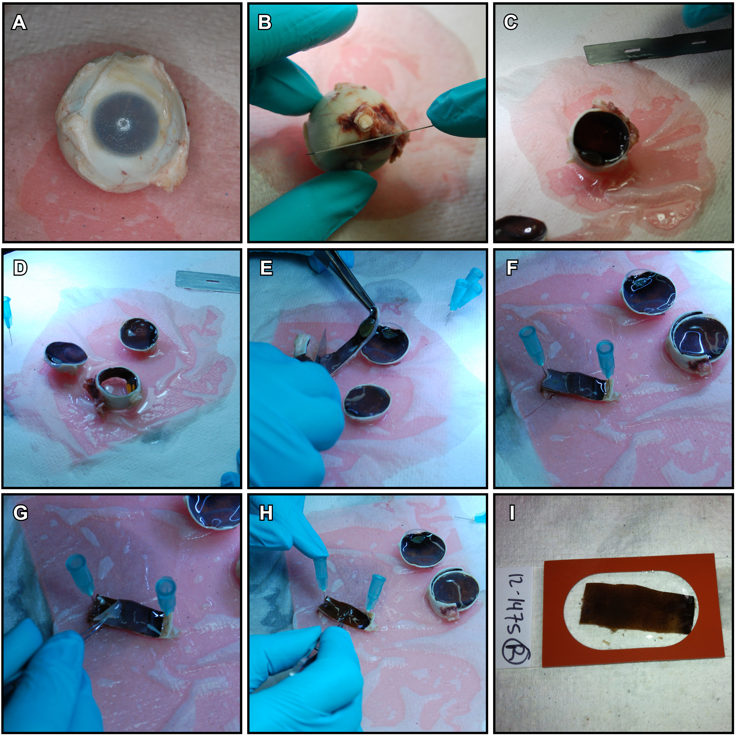
 Figure 1 of
Bhatia, Mol Vis 2016; 22:898-916.
Figure 1 of
Bhatia, Mol Vis 2016; 22:898-916.