Figure 3. TF promotes VEGF expression in ARPE-19 cells. A: The vascular endothelial growth factor (VEGF) expression detected with western blotting in ARPE-19 cells following tissue
factor (TF) siRNA transfection after 48 h. B: Histogram shows densitometric analysis of the average levels for TF and VEGF. Glyceraldehyde 3-phosphate dehydrogenase (GAPDH)
was used as an internal control. Cells transfected with scrambled siRNA is used as negative control. C: Western blot analysis of VEGF expression in ARPE-19 cells after pEGFP-N3-hTF plasmid transfection after 48 h. D: Histogram shows densitometric analysis of the average levels for TF and VEGF. GAPDH was used as an internal control. Cells
transfected with empty plasmid is used as negative control. * p<0.01, TF siRNA-treated group versus control groups.
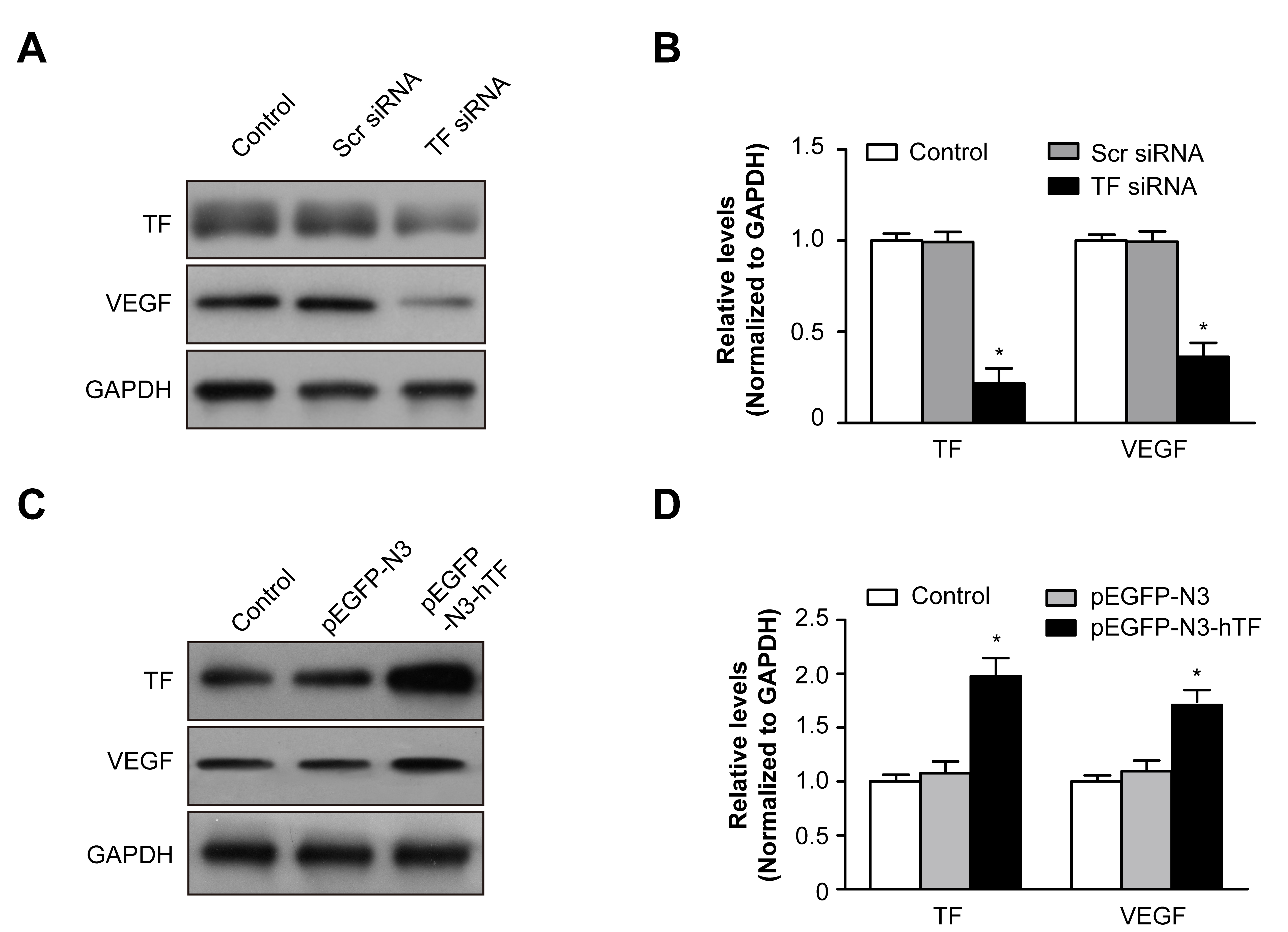
 Figure 3 of
Wang, Mol Vis 2016; 22:886-897.
Figure 3 of
Wang, Mol Vis 2016; 22:886-897.