Figure 3. Confocal images reveal that two different isozymes of hexokinase, HK-1 and HK-2, have distinct compartmental and laminar distribution
in the adult mouse retina. A: The anti-HK-1 antibody selectively and strongly labels the INL. B: High magnification of the OPL from retinas double-labeled for HK-1 and vesicular glutamate transporter 1 (VGLUT1) show no
colocalization. C: A high-magnification image of the OPL from retinas double-labeled for HK-1 and protein kinase C alpha (PKCα) shows strong
colocalization. D: Retinas double-labeled for HK-1 and glutamine synthetase (GS) show weak colocalization in the MGC somas. E: The anti-HK-2 antibody differentially labels the outer and inner retina. The anti-HK-2 antibody intensely labels the ISs
and the OPL. F: A high-magnification image of the OPL double-labeled for HK-2 and VGLUT1 shows colocalization. G: HK-2 and PKCα do not colocalize. H: HK-2 and calbindin colocalize in horizontal cell axonal processes with rod terminals (white arrows) and in dendritic processes
with cone terminals (white arrowheads). I: The outer retina of the neural retina leucine zipper–green fluorescent protein (Nrl-GFP) transgenic mice (pseudocolored in red) labeled for HK-2 shows colocalization in the rod IS and spherules (yellow-orange
pixels) and HK-2 expression in the cone OSs and ISs (green pixels: black arrows) and pedicles (green pixels). J: High-magnification image of the OS-IS from Nrl-GFP transgenic mice (pseudocolored in red). K: High-magnification image of Nrl-GFP retina colabeled for HK-2 shows colocalization in the rod ISs and spherules (yellow-orange pixels) and localization of
HK-2 in the cone OSs and ISs (green pixels) and somas in the ONL (green pixels: white arrowheads). GCL = ganglion cell layer,
INL = inner nuclear layer, IPL = inner plexiform layer, IPL-a = IPL sublamina-a, IPL-b = sublamina-b, ISs = inner segments,
ONL = outer nuclear layer, OPL = outer plexiform layer, OSs = outer segments. A, D, E, and I, scale bar = 40 µm. B–C and F–H,
scale bar = 20 µm. J and K, the scale bar = 10 μm.
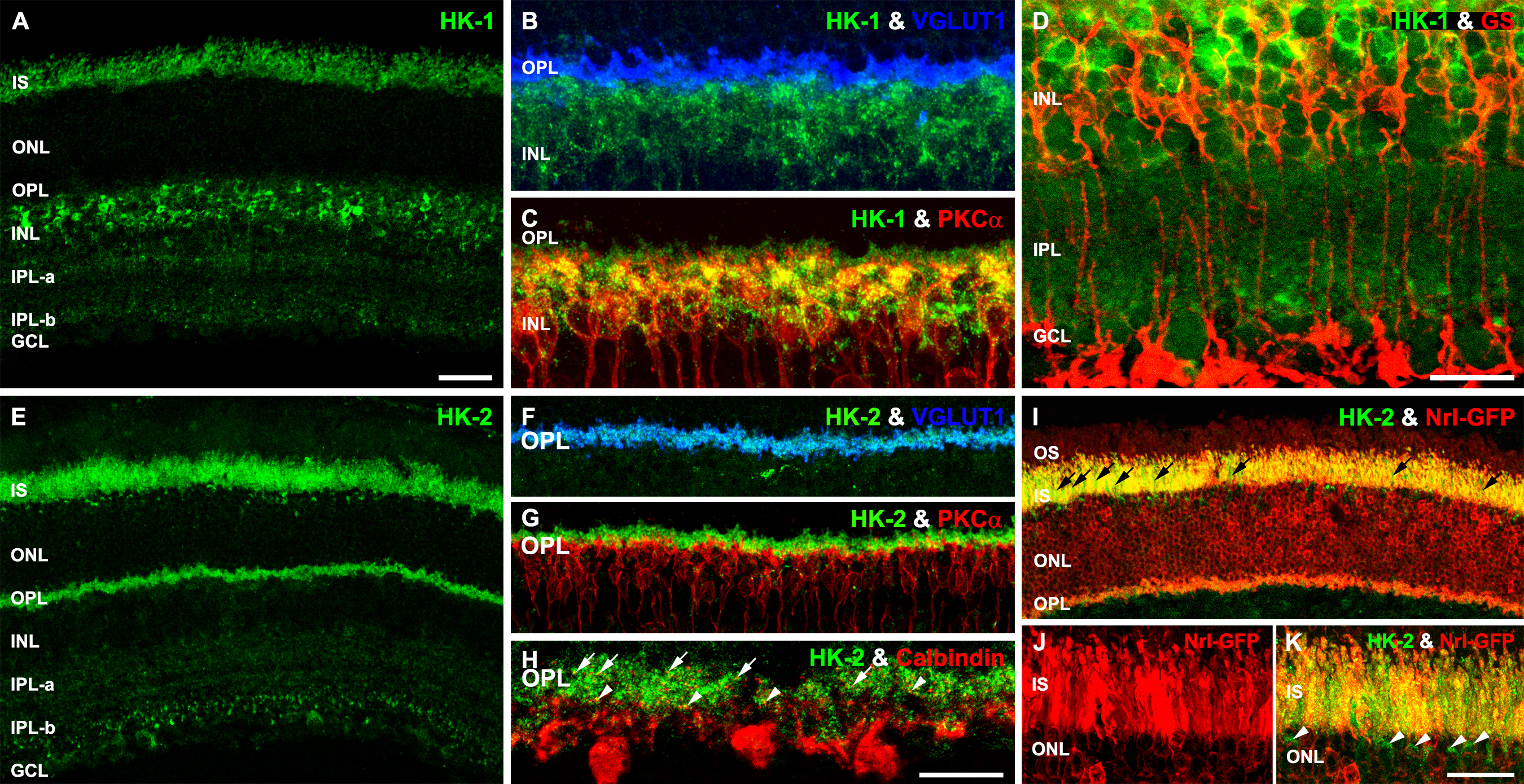
 Figure 3 of
Rueda, Mol Vis 2016; 22:847-885.
Figure 3 of
Rueda, Mol Vis 2016; 22:847-885.