Figure 2. The relative location of the rod and cone outer segments, and myoid and ellipsoid regions of inner segments in the adult mouse
retina are presented: electron and confocal microscopy images. Note that the cone OSs are located in the same layer as the
rod inner segments (RISs). A: Low-magnification electron micrograph of a longitudinal section of the entire photoreceptor layer. Note the presence of
cone OSs in the rod IS layer (white arrows) and the perinuclear mitochondria of the cones (white arrowheads). B: All compartments of the rods in the neural retina leucine zipper–green fluorescent protein (Nrl-GFP) transgenic mouse are fluorescent for GFP. C: Cytochrome oxidase subunit IV (COX IV) immunoreactivity (IR) colocalizes in the mitochondria-rich rod IS ellipsoid region
and the OPL of Nrl-GFP transgenic mice (yellow-orange pixels). White arrowheads identify the cone perinuclear mitochondria. D: High-magnification image of Nrl-GFP transgenic mouse retina shows cone S-opsin-IR in the rod IS layer. E: High magnification of a retina double-labeled with COX IV and cone arrestin (M-CAr) shows cones (green pixels) in the rod
IS ellipsoid region. White arrowheads identify the colabeled cone perinuclear mitochondria. A, scale bar = 10 μm. B and C, scale bar = 40 μm. D and E, scale bar = 20 μm. ELM = external limiting membrane, ellip = ellipsoid, ISs = inner segments, myo = myoid, ONL = outer nuclear
layer, OPL = outer plexiform layer, OSs = outer segments.
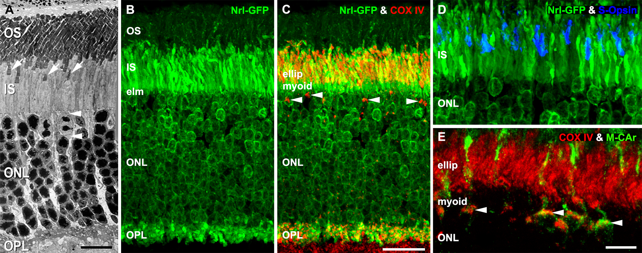
 Figure 2 of
Rueda, Mol Vis 2016; 22:847-885.
Figure 2 of
Rueda, Mol Vis 2016; 22:847-885.