Figure 15. Confocal images illustrate that the creatine kinase isozyme found in muscle and the brain (CK-M) is distributed in cellular
regions of the retina where endoplasmic reticulum proteins are located. A: A transverse section of mouse gastrocnemius immunolabeled for CK-M. The inset shows a longitudinal section of the gastrocnemius
immunolabeled for CK-M and stained with the nuclear dye DRAQ5. B: A longitudinal section of gastrocnemius triple-labeled for CK-M, COX IV, and DRAQ5. The inset shows a high-magnification
image of a transverse section of the gastrocnemius double-labeled for CK-M and COX IV. C: A sagittal section of the mouse frontal cortex immunolabeled for CK-M. The inset shows a high magnification image of cortical
cells labeled for CK-M and stained with DRAQ5. D: A higher magnification image of frontal cortex triple-labeled for CK-M, COX IV, and DRAQ5. E: Retina immunolabeled for CK-M. F: Outer retina double-labeled for CK-M and VGLUT1. G: Outer retina double-labeled for SERCA3 and VGLUT1. H and I: High-magnification images of the outer retina double-labeled for CK-M and COX IV. J: Retina double-labeled for CK-M and PKCα. COX IV = cytochrome c oxidase subunit IV, GCL = ganglion cell layer, INL = inner
nuclear layer, IPL = inner plexiform layer, ISs = inner segments, ONL = outer nuclear layer, OPL = outer plexiform layer,
PKCα = protein kinase C α, VGLUT1 = vesicular glutamate transporter 1. A–C, E–G and J, scale bar = 40 µm. D, scale bar = 10
µm. H and I, scale bar = 20 µm.
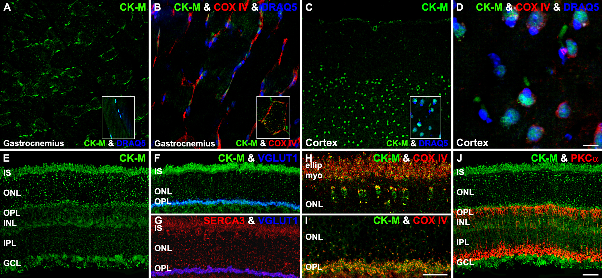
 Figure 15 of
Rueda, Mol Vis 2016; 22:847-885.
Figure 15 of
Rueda, Mol Vis 2016; 22:847-885.