Figure 14. Confocal images show selective expression of the ubiquitous mi-CK in mouse retina. A: Retinas immunolabeled for mitochondrial creatine kinase (mi-CK) show that the ISs, distal ONL, OPL, GCL, and NFL/MGC end-feet
are labeled. B: Retinas double-label with mi-CK and GS show moderate mi-CK-IR in the MGC end-feet. C: Retinas double-labeled with mi-CK and COX IV show that the cone ISs and the perinuclear mitochondria of cone somas in the
distal ONL are intensely labeled while rod inner segments (RISs) and spherules show weak to moderate mi-CK-IR. COX IV = cytochrome
c oxidase subunit IV, GCL = ganglion cell layer, GS = glutamine synthetase, IPL = inner plexiform layer, ISs = inner segments,
MGC = Müller glial cell, NFL = nerve fiber layer, ONL = outer nuclear layer, OPL = outer plexiform layer. A and B, scale bar
= 40 µm. C, scale bar = 20 µm.
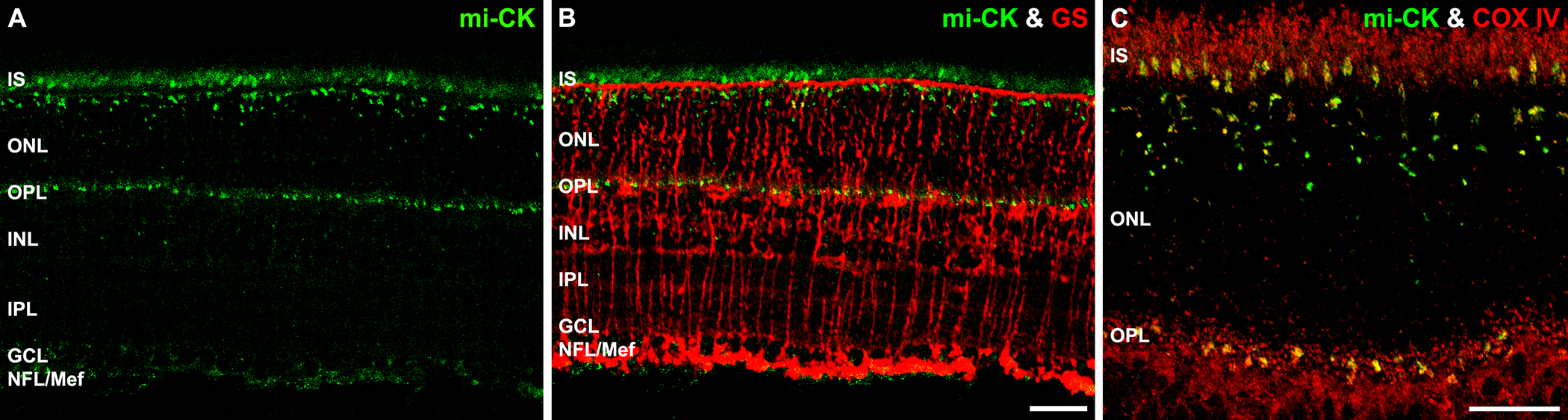
 Figure 14 of
Rueda, Mol Vis 2016; 22:847-885.
Figure 14 of
Rueda, Mol Vis 2016; 22:847-885.