Figure 12. Confocal images show that two functionally distinct isozymes of adenylate kinase (AK1 and AK2) are differentially expressed
throughout the retina. A: AK1 is selectively expressed in the inner retina. B: Retinas double-labeled for AK1 and GLAST show no colocalization. C: A high-magnification image of the retina reveals that AK1 does not colocalize with either CHX10 or VGLUT1. D–F: High magnification images of the inner retina double-labeled for AK1 and three antibodies for different types of amacrine
cells. D: AK1 and GABA colocalize in weakly GABAergic-IR cells in the INL and the GCL (white arrows). E: AK1 and ChAT do not colocalize in cholinergic amacrine cells or the two cholinergic strata in the IPL. F: AK1 and disabled 1 (DAB1) colocalize in distal amacrine cells (white arrows). G: Retina immunolabeled for AK2. H: Retinas double-labeled for AK2 and VGLUT1 show colocalization in the OPL and the IPL (aquamarine pixels). I: Retinas double-labeled for AK2 and GLAST show extensive colocalization (yellow-orange pixels). ChAT = choline acetyltransferase,
GCL = ganglion cell layer, GLAST = glutamate-aspartate transporter, INL = inner nuclear layer, IPL = inner plexiform layer,
ISs = inner segments, ONL = outer nuclear layer, OPL = outer plexiform layer, VGLUT1 = vesicular glutamate transporter 1.
A, B, D, and F–I, scale bar = 40 µm. C, scale bar = 20 µm. E, scale bar = 20 µm.
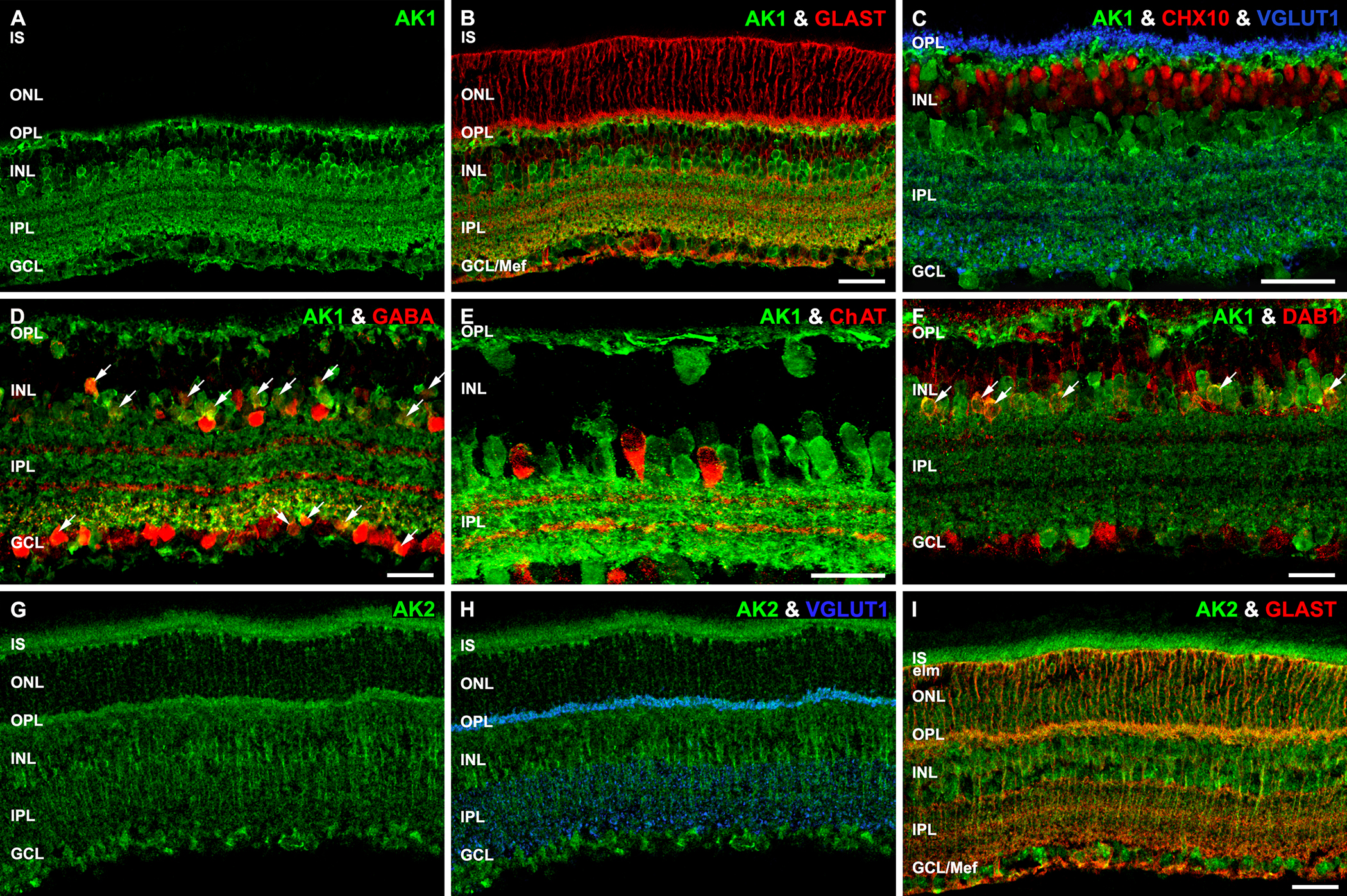
 Figure 12 of
Rueda, Mol Vis 2016; 22:847-885.
Figure 12 of
Rueda, Mol Vis 2016; 22:847-885.