Figure 11. Confocal images show that NDPK is widely expressed throughout the retina. A: Retina immunolabeled for nucleoside diphosphate kinase isoform A (NDPK). B: Retinas double-labeled for NDPK and the glial high-affinity glutamate-aspartate transporter (GLAST) show colocalization
in all MGC regions: the ELM, distal and proximal processes, soma, and end-feet (yellow-orange pixels). C: A higher-magnification image shows colocalization of NDPK and GS in the distal and proximal MGC processes (yellow-orange
pixels). D: A high-magnification image of a retina double-labeled for NDPK and VGLUT1 reveals greater colocalization in the OPL than
in the IPL (aquamarine pixels). COX IV-IR = cytochrome oxidase IV immunoreactivity, ELM = external limiting membrane, GCL
= ganglion cell layer, GS = glutamine synthetase, INL = inner nuclear layer, IPL = inner plexiform layer, IPL-a = IPL sublamina-a,
IPL-b = IPL sublamina-b, ISs = inner segments, MGC = Müller glial cell, ONL = outer nuclear layer, OPL = outer plexiform layer,
VGLUT1 = vesicular glutamate transporter 1. A and B, scale bar = 40 µm. C and D, scale bar = 20 µm.
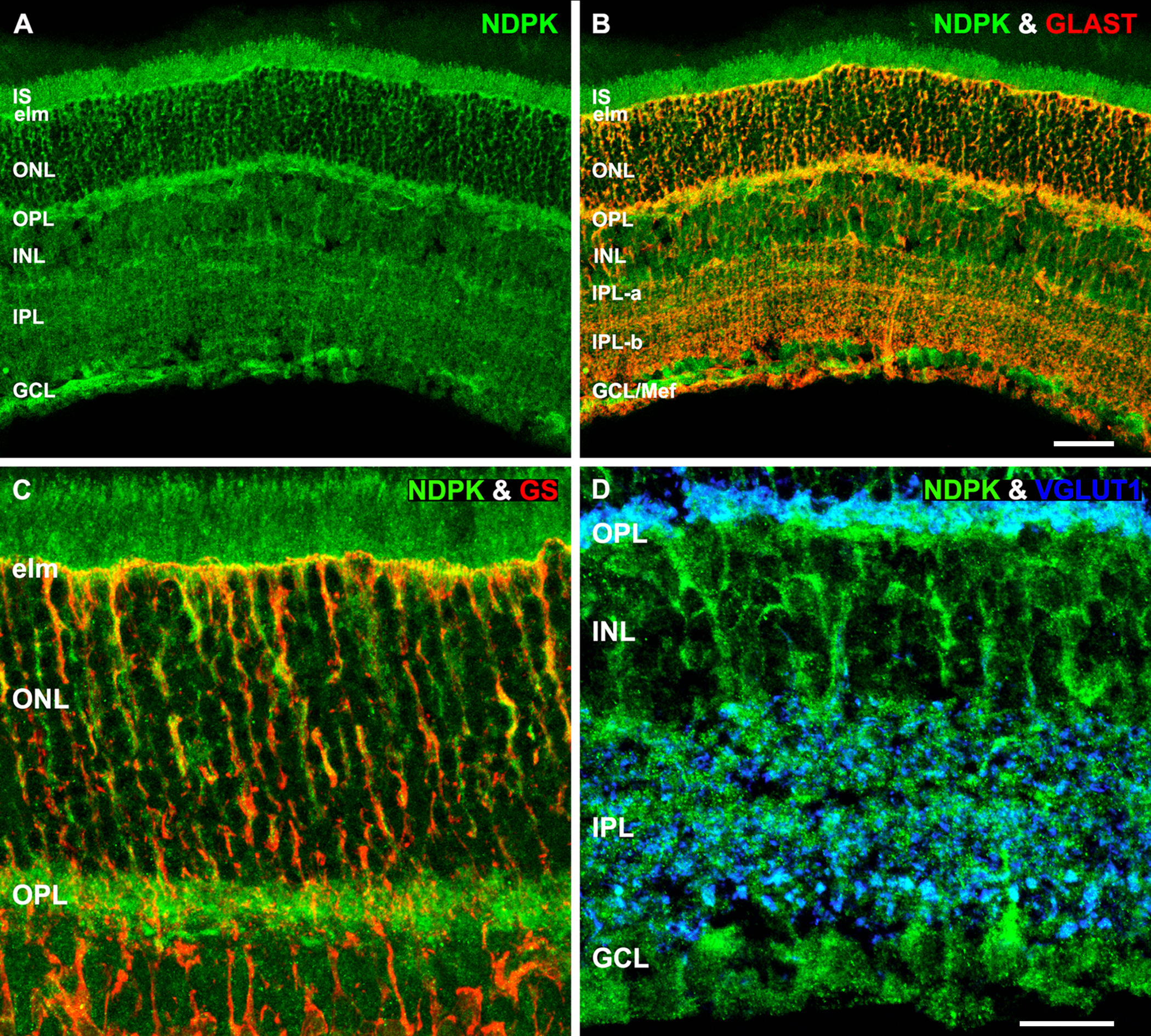
 Figure 11 of
Rueda, Mol Vis 2016; 22:847-885.
Figure 11 of
Rueda, Mol Vis 2016; 22:847-885.