Figure 3. Fasudil suppresses the LPS-stimulated phosphorylation of p38 MAPK in RMG cells. A: The protein levels of phosphor-p38 mitogen-activated protein kinase (p-p38-MAPK) and phosphor-p38-MAPK (p-p38-MAPK) in lipopolysaccharide
(LPS) stimulation and/or fasudil-treated retinal microglial (RMG) cells. The cells were pretreated with SB203580 (20 mM) for
30 min and incubated in the presence or absence of fasudil (50 μM) for 6 h in LPS-stimulated RMG cells. B: The relative protein band intensities were quantified with densitometric analyses and normalized to β-actin, p38-MAPK, and
p-p38-MAPK. Values represent the means ± standard deviation (SD) of three independent experiments performed in triplicate.
#p<0.05 compared with untreated cells, **p<0.05 compared with LPS-stimulated cells.
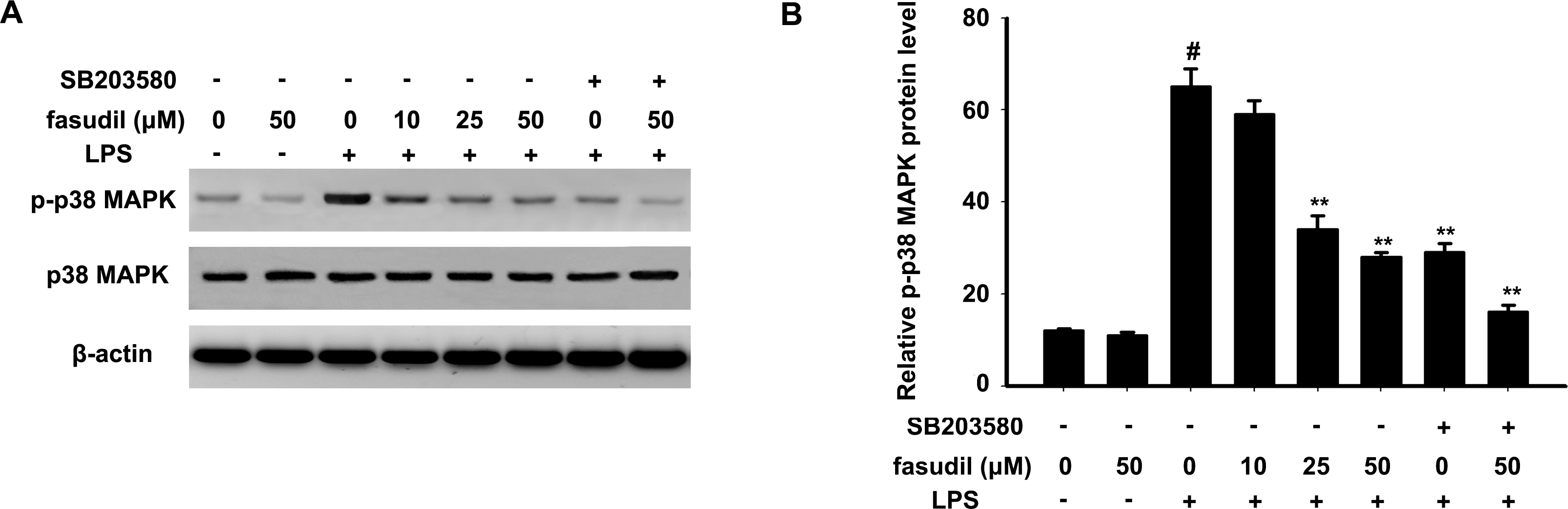
 Figure 3 of
Xu, Mol Vis 2016; 22:836-846.
Figure 3 of
Xu, Mol Vis 2016; 22:836-846.