Figure 1. Primary RMG cells were identified with immunofluorescence staining of OX42. A: OX42 (red). B: Nuclear Hoechst staining (blue). C: The purple color visualized in the merged images represents the colocalization of OX42 with nuclear Hoechst staining. Scale
bars are equivalent to 25 μm.
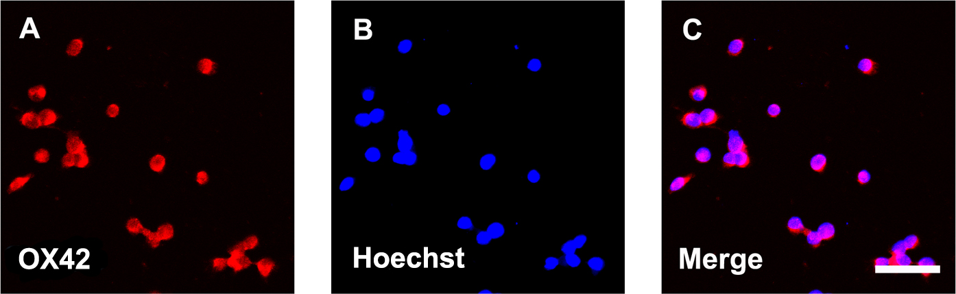
 Figure 1 of
Xu, Mol Vis 2016; 22:836-846.
Figure 1 of
Xu, Mol Vis 2016; 22:836-846.