Figure 3. Scleral expression of thrombospondin and HINT1. Immunostaining for thrombospondin-1 (TSP-1; red) counterstained with 4’,6-diamidino-2-phenylindole
(DAPI; blue) shows labeling comparable to the mean increase in the masked scleral grading (see text). The sclera is marked
with a bracket in each panel. A: Control. B: Increased labeling in the 3-day glaucoma sclera, choroid, and retina (CD1 mice). Scale bar=50 µm. Lower pair shows immunostaining
for HINT1 (red), sclera labeling in the (C) CD1 control and increased labeling in the glaucoma tissue (D). DAPI counterstain (blue in A, B; green in C, D). Scale bar=25 µm.
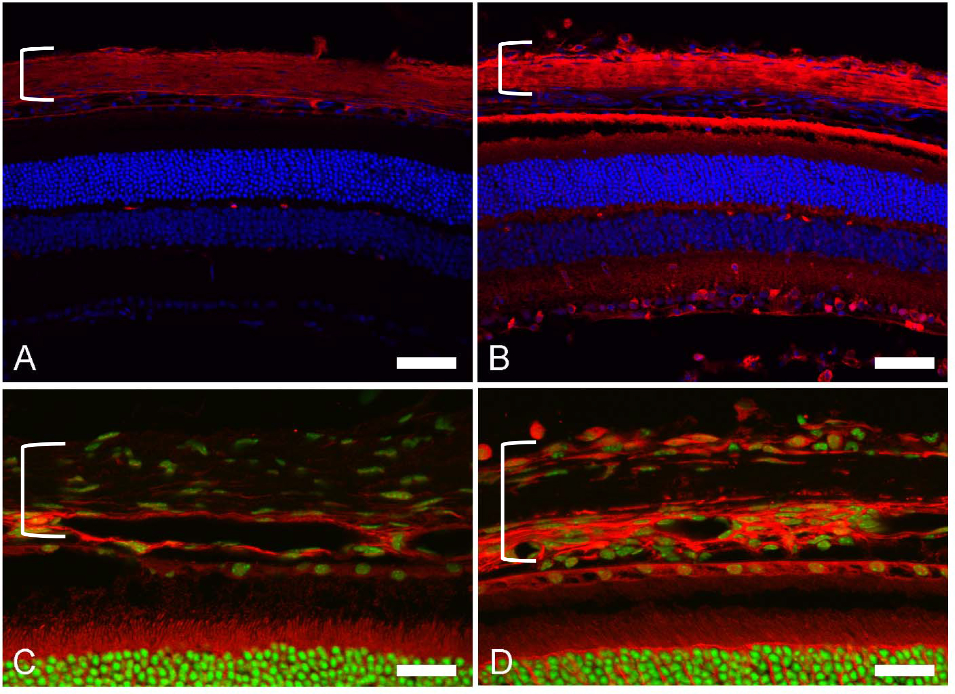
 Figure 3 of
Oglesby, Mol Vis 2016; 22:82-99.
Figure 3 of
Oglesby, Mol Vis 2016; 22:82-99.